. 2021 Jun 17;11(6):1812.
doi: 10.3390/ani11061812.

Effects of Orexin B on Swine Granulosa and Endothelial Cells

Affiliations

Effects of Orexin B on Swine Granulosa and Endothelial Cells

Francesca Grasselli et al. Animals (Basel). .

Abstract

In addition to the well-known central modulatory role of orexins, we recently demonstrated a peripheral involvement in swine granulosa cells for orexin A and in adipose tissue for orexin B (OXB). The aim of present research was to verify immunolocalization of OXB and its potential role in modulating the main features of swine granulosa cells. In particular, we explored the effects on granulosa cell proliferation (through the incorporation of bromodeoxyuridine), cell metabolic activity (as indirect evaluation by the assessment of ATP), steroidogenic activity (by immunoenzymatic examination) and redox status (evaluating the production of superoxide anion by means of the WST test, production of nitric oxide through the use of the Griess test and the non-enzymatic reducing power by FRAP test). Our data point out that OXB does not modify granulosa cell growth, steroidogenesis and superoxide anion generation. On the contrary, the peptide stimulates (p < 0.05) nitric oxide output and non-enzymatic reducing power. Since new vessel growth is crucial for ovarian follicle development, a further aim of this study was to explore the expression of prepro-orexin and the effects of OXB on swine aortic endothelial cells. We found that the peptide is ineffective in modulating cell growth, while it inhibits redox status parameters. In addition, we demonstrated a stimulatory effect on angiogenesis evaluated in fibrin gel angiogenesis assay. Taken together, OXB appears to be potentially involved in the modulation of redox status in granulosa and endothelial cells and we could argue an involvement of the peptide in the follicular angiogenic events.

Keywords: angiogenesis; estradiol 17 β; ovarian follicle; oxidative stress; progesterone.


Conflict of interest statement

The authors declare no conflict of interest.

Figures

Figure 1
Figure 1
(A) Cytoplasmatic positivity for orexin B in granulosa cells (20X IHC). Scale bar = 100 µm. (B) Negative control.
Figure 2
Figure 2
Effect of the 48 h treatment with orexin B (OXB; 0.1, 1 and 10 nM) on swine granulosa cell viability, evaluated by MTT test (A), and on granulosa cell proliferation evaluated using 5-bromo-2′-deoxyuridine (BrdU) incorporation assay test (B). Data, expressed as milliAbsorbance (mAbs), represent the mean ± SEM of six replicates/treatment repeated in five different experiments.
Figure 3
Figure 3
Effect of the 48 h treatment with orexin B (OXB; 0.1, 1 and 10 nM) on swine granulosa cell estradiol 17β (E2) and progesterone (P4) production (panels (A) and (B), respectively) evaluated by ELISA tests. Data, expressed as ng/mL, represent the mean ± SEM of six replicates/treatment repeated in five different experiments.
Figure 4
Figure 4
Effect of the 48 h treatment with orexin B (OXB; 0.1, 1 and 10 nM) on swine granulosa cell superoxide anion generation (O2) using WST assay (A), nitric oxide (NO) production using Griess assay (B) and non-enzymatic scavenging activity using FRAP assay (C). Data, expressed as milliAbsorbance (mAbs), represent the mean ± SEM of six replicates/treatment repeated in five different experiments. In each panel, different letters on the bars indicate a significant difference (p < 0.05) among treatments as calculated by ANOVA and Scheffè’s F test.
Figure 5
Figure 5
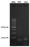
Prepro-orexin (PPO) and actin (ACT) expression in swine aortic endothelial cells (AOC). PPO expression was evaluated by nested PCR; the presence of PPO amplicon demonstrates gene expression but does not provide any information in comparison to ACT expression.
Figure 6
Figure 6
Effect of the 48 h treatment with orexin B (OXB; 10 nM) on swine aortic endothelial cells’ viability, evaluated by MTT test (A) and on granulosa cell proliferation evaluated using 5-bromo-2′-deoxyuridine (BrdU) incorporation assay test (B).
Figure 7
Figure 7
Effect of the 48 h treatment with orexin B (OXB; 10 nM) on swine aortic endothelial cells’ superoxide anion generation (O2) using WST assay (A), nitric oxide (NO) production using Griess assay (B) and non-enzymatic scavenging activity using FRAP assay (C). Data, expressed as milliAbsorbance (mAbs), represent the mean ± SEM of six replicates/treatment repeated in five different experiments. In each panel, different letters on the bars indicate a significant difference (p < 0.05) among treatments as calculated by ANOVA and Scheffè’s F test.
Figure 8
Figure 8
Effect of the 48 h treatment with OXB (10 nM) on aortic endothelial cell (AOC) growth in fibrin gels. Panel (A): data, expressed as pixels, represent the mean ± SEM of six replicates/treatment repeated in five different experiments. Panel (B): exemplary images of the AOC growing on collagen-coated MC beads in fibrin gels, both after 48 h of treatment with OXB (10 nM).
Figure 9
Figure 9
Effect of the 72 h treatment with OXB (10 nM) on aortic endothelial cell (AOC) growth in fibrin gels. Panel (A): data, expressed as pixels, represent the mean ± SEM of six replicates/treatment repeated in five different experiments. Different letters on the bars indicate a significant difference (p < 0.001) among treatments as calculated by ANOVA and Scheffè’s F test. Panel (B): exemplary images of the AOC growing on collagen-coated MC beads in fibrin gels both after 72 h of treatment with OXB (10 nM).

References

    1. de Lecea L., Kilduff T.S., Peyron C., Gao X., Foye P.E., Danielson P.E., Fukuhara C., Battenberg E.L., Gautvik V.T., Bartlett F.S., 2nd, et al. The hypocretins: Hypothalamus-specific peptides with neuroexcitatory activity. Proc. Natl. Acad. Sci. USA. 1998;95:322–327. doi: 10.1073/pnas.95.1.322. - DOI - PMC - PubMed
    1. Sakurai T., Amemiya A., Ishii M., Matsuzaki I., Chemelli R.M., Tanaka H., Williams S.C., Richardson J.A., Kozlowski G.P., Wilson S., et al. Orexins and orexin receptors: A family of hypothalamic neuropeptides and G protein-coupled receptors that regulate feeding behavior. Cell. 1998;92:573–585. doi: 10.1016/S0092-8674(00)80949-6. - DOI - PubMed
    1. Liu L., Wang Q., Liu A., Lan X., Huang Y., Zhao Z., Jie H., Chen J., Zhao Y. Physiological Implications of Orexins/Hypocretins on Energy Metabolism and Adipose Tissue Development. ACS Omega. 2019;5:547–555. doi: 10.1021/acsomega.9b03106. - DOI - PMC - PubMed
    1. Sakurai T. The neuralcircuit of orexin (hypocretin): Maintaining sleep and wakefulness. Nat. Rev. Neurosci. 2007;8:171–181. doi: 10.1038/nrn2092. - DOI - PubMed
    1. Chieffi S., Carotenuto M., Monda V., Valenzano A., Villano I., Precenzano F., Tafuri D., Salerno M., Filippi N., Nuccio F., et al. Orexin System: The Key for a Healthy Life. Front. Physiol. 2017;8:357. doi: 10.3389/fphys.2017.00357. - DOI - PMC - PubMed

LinkOut - more resources