. 2021 Jun 3;5(2):27.
doi: 10.3390/vision5020027.

Ocular Surface Microbiota in Contact Lens Users and Contact-Lens-Associated Bacterial Keratitis

Affiliations

Ocular Surface Microbiota in Contact Lens Users and Contact-Lens-Associated Bacterial Keratitis

Jasmine Andersson et al. Vision (Basel). .

Abstract

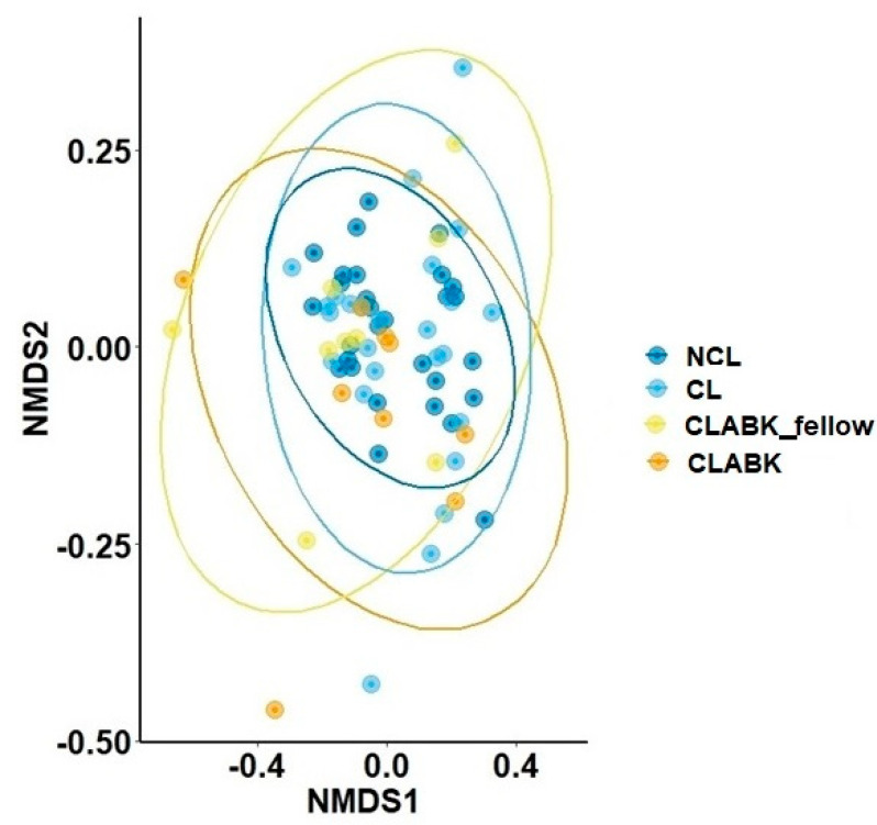
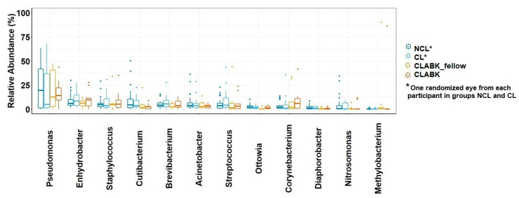
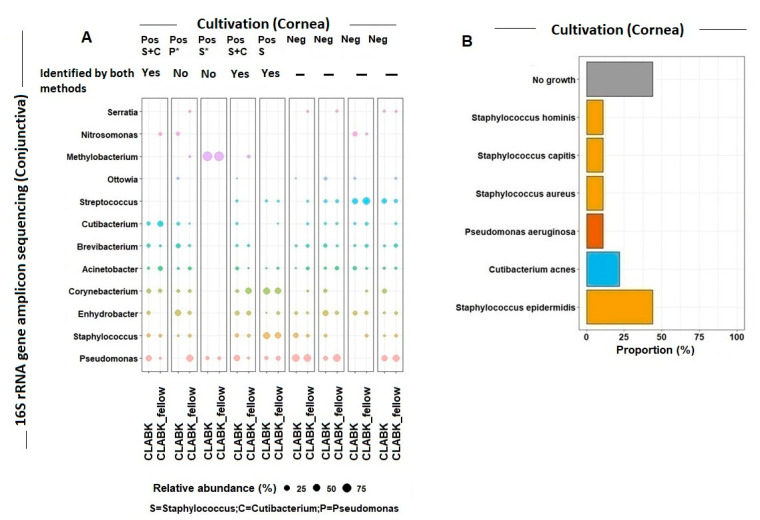
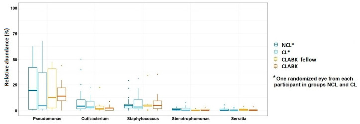
Our objectives were to investigate whether the conjunctival microbiota is altered by contact lens wear and/or bacterial keratitis and to explore the hypothesis that commensals of conjunctival microbiota contribute to bacterial keratitis. Swab samples from both eyes were collected separately from the inferior fornix of the conjunctiva of non-contact-lens users (nparticipants = 28) and contact lens users (nparticipants = 26) and from patients with contact-lens-associated bacterial keratitis (nparticipants = 9). DNA from conjunctival swab samples was analyzed with 16S rRNA gene amplicon sequencing. Pathogens from the corneal infiltrates were identified by cultivation. In total, we identified 19 phyla and 283 genera; the four most abundant genera were Pseudomonas, Enhydrobacter, Staphylococcus, and Cutibacterium. Several pathogens related to bacterial keratitis were identified in the conjunctival microbiota of the whole study population, and the same bacteria were identified by both methods in the conjunctiva and cornea for four patients with contact-lens-associated bacterial keratitis. The overall conjunctival microbiota profile was not altered by contact lens wear or bacterial keratitis; thus, it does not appear to contribute to the development of bacterial keratitis in contact lens users. However, in some individuals, conjunctival microbiota may harbor opportunistic pathogens causing contact-lens-associated bacterial keratitis.

Keywords: 16S rRNA gene amplicon sequencing; bacterial keratitis; conjunctival microbiota; contact lenses; contact-lens-associated bacterial keratitis; cultivation; ocular surface microbiota; opportunistic pathogens.


Conflict of interest statement

The authors declare no conflict of interest.

Figures

Figure 1
Figure 1
Comparison of the bacterial composition of ocular surface microbiota using the Bray–Curtis dissimilarity index for the study population. The bacterial composition was compared at the amplicon sequence variant (ASV) level and visualized in a two-dimensional plot with nonmetric multidimensional scaling (NMDS). Conjunctival samples from both eyes of the patients with contact-lens-associated bacterial keratitis (eye with bacterial keratitis (CLABK) and the eye without bacterial keratitis (CLABK_fellow)) were compared with one randomized eye for each participant in the following groups: contact lens users without bacterial keratitis (CL) and non-contact-lens users (NCL). The bacterial composition was not significantly different between groups (pairwise comparison with PERMANOVA); NCL vs. CL p = 1.0; NCL vs. CLABK_fellow p = 0.49; NCL vs. CLABK p = 0.35; CL vs. CLABK_fellow p = 1.0; CL vs. CLABK p = 0.82. Stress ratio = 0.14.
Figure 2
Figure 2
Relative abundance (%) of the 12 most common genera of ocular surface microbiota in the whole study population. Boxplots for relative abundance with medians and interquartile range (IQR) are shown for non-contact-lens users (NCL), contact lens users without bacterial keratitis (CL), fellow eye of patients with contact-lens-associated bacterial keratitis (CLABK_fellow) and for eyes with bacterial keratitis (CLABK). Data from one randomized eye of each participant in the NCL and CL groups were compared with data from CLABK_fellow and CLABK. No significant difference was demonstrated with the Kruskal–Wallis test (padj > 0.05) for NCL vs. CL/CLABK_fellow or for NCL vs. CL/CLABK.
Figure 3
Figure 3
(A) 16S rRNA gene amplicon sequencing versus cultivation of the ocular surface microbiota. For contact lens users with bacterial keratitis (i.e., CLABK = eye with bacterial keratitis; CLABK_fellow = eye without bacterial keratitis), the relative abundance for the most common genera identified in the conjunctiva using 16S rRNA gene amplicon sequencing was compared with the cultivation results from the sampling of the corneal infiltrates. Five cultivations were positive for growth (Pos), defined as ≥one bacterial species, and four cultivations were without growth (Neg). The potential bacterial pathogens of the cornea were identified at the genus level as Staphylococcus (S), Cutibacterium (C), and Pseudomonas (P). For four out of the five positive cultures, the same bacteria at the genus level were detected in the CLABK samples with 16S rRNA gene amplicon sequencing and by cultivation. (The relative abundance for Staphylococcus was ≤1% in one CLABK sample and therefore not shown in the figure (marked with *)). Pseudomonas was identified by cultivation in one patient; however, the relative abundance was 0% in the CLABK sample using 16S rRNA gene amplicon sequencing. Minimum relative abundance for the shown genera was 1%. (B) Cultivation results of the corneal infiltrates in the CLABK samples. Six bacterial species were identified, and Staphylococcus epidermidis was the most frequently cultivated potential pathogen with growth in four out of five patients (80%). One to three bacterial species were cultivated per patient, while cultivations for four out of nine patients (44%) showed no growth.
Figure 4
Figure 4
The relative abundance of five known bacterial pathogens to bacterial keratitis. No significant difference of the relative abundance of these five genera was demonstrated for the comparison between non-contact-lens users (NCL) vs. contact lens users (CL)/fellow eye of patient with contact-lens-associated bacterial keratitis (CLABK_fellow) or for the comparison between NCL/CL vs. eyes with bacterial keratitis (CLABK) (Kruskal–Wallis test, p > 0.05).

References

    1. Ung L., Bispo P.J.M., Shanbhag S.S., Gilmore M.S., Chodosh J. The persistent dilemma of microbial keratitis: Global burden, diagnosis, and antimicrobial resistance. Surv. Ophthalmol. 2019;64:255–271. doi: 10.1016/j.survophthal.2018.12.003. - DOI - PMC - PubMed
    1. Green M., Apel A., Stapleton F. Risk factors and causative organisms in microbial keratitis. Cornea. 2008;27:22–27. doi: 10.1097/ICO.0b013e318156caf2. - DOI - PubMed
    1. Austin A., Lietman T., Rose-Nussbaumer J. Update on the Management of Infectious Keratitis. Ophthalmology. 2017;124:1678–1689. doi: 10.1016/j.ophtha.2017.05.012. - DOI - PMC - PubMed
    1. Bourcier T., Thomas F., Borderie V., Chaumeil C., Laroche L. Bacterial keratitis: Predisposing factors, clinical and microbiological review of 300 cases. Br. J. Ophthalmol. 2003;87:834–838. doi: 10.1136/bjo.87.7.834. - DOI - PMC - PubMed
    1. Galdiero M., Petrillo F., Pignataro D., Lavano M.A., Santella B., Folliero V., Zannella C., Astarita C., Gagliano C., Franci G., et al. Current evidence on the ocular surface microbiota and related diseases. Microorganisms. 2020;8:1–13. - PMC - PubMed

LinkOut - more resources