

Comparison of Primary Virus Isolation in Pulmonary Alveolar Macrophages and Four Different Continuous Cell Lines for Type 1 and Type 2 Porcine Reproductive and Respiratory Syndrome Virus
- PMID: 34205087
- PMCID: PMC8229515
- DOI: 10.3390/vaccines9060594
Comparison of Primary Virus Isolation in Pulmonary Alveolar Macrophages and Four Different Continuous Cell Lines for Type 1 and Type 2 Porcine Reproductive and Respiratory Syndrome Virus
Abstract
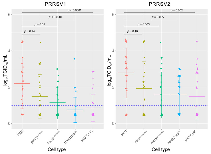
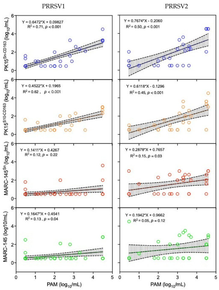
Porcine Reproductive and Respiratory Syndrome Virus (PRRSV) has a highly restricted cellular tropism. In vivo, the virus primarily infects tissue-specific macrophages in the nose, lungs, tonsils, and pharyngeal lymphoid tissues. In vitro however, the MARC-145 cell line is one of the few PRRSV susceptible cell lines that are routinely used for in vitro propagation. Previously, several PRRSV non-permissive cell lines were shown to become susceptible to PRRSV infection upon expression of recombinant entry receptors (e.g., PK15Sn-CD163, PK15S10-CD163). In the present study, we examined the suitability of different cell lines as a possible replacement of primary pulmonary alveolar macrophages (PAM) cells for isolation and growth of PRRSV. The susceptibility of four different cell lines (PK15Sn-CD163, PK15S10-CD163, MARC-145, and MARC-145Sn) for the primary isolation of PRRSV from PCR positive sera (both PRRSV1 and PRRSV2) was compared with that of PAM. To find possible correlations between the cell tropism and the viral genotype, 54 field samples were sequenced, and amino acid residues potentially associated with the cell tropism were identified. Regarding the virus titers obtained with the five different cell types, PAM gave the highest mean virus titers followed by PK15Sn-CD163, PK15S10-CD163, MARC-145Sn, and MARC-145. The titers in PK15Sn-CD163 and PK15S10-CD163 cells were significantly correlated with virus titers in PAM for both PRRSV1 (p < 0.001) and PRRSV2 (p < 0.001) compared with MARC-145Sn (PRRSV1: p = 0.22 and PRRSV2: p = 0.03) and MARC-145 (PRRSV1: p = 0.04 and PRRSV2: p = 0.12). Further, a possible correlation between cell tropism and viral genotype was assessed using PRRSV whole genome sequences in a Genome-Wide-Association Study (GWAS). The structural protein residues GP2:187L and N:28R within PRRSV2 sequences were associated with their growth in MARC-145. The GP5:78I residue for PRRSV2 and the Nsp11:155F residue for PRRSV1 was linked to a higher replication on PAM. In conclusion, PK15Sn-CD163 and PK15S10-CD163 cells are phenotypically closely related to the in vivo target macrophages and are more suitable for virus isolation and titration than MARC-145/MARC-145Sn cells. The residues of PRRSV proteins that are potentially related with cell tropism will be further investigated in the future.
Keywords: MARC-145; PK15S10-CD163; PK15Sn-CD163; PRRSV; macrophages; virus isolation/production.
Conflict of interest statement
The authors declare no conflict of interest.
Figures
Similar articles
-
Susceptible cell lines for the production of porcine reproductive and respiratory syndrome virus by stable transfection of sialoadhesin and CD163.BMC Biotechnol. 2010 Jun 29;10:48. doi: 10.1186/1472-6750-10-48. BMC Biotechnol. 2010. PMID: 20587060 Free PMC article.
-
Preferential use of Siglec-1 or Siglec-10 by type 1 and type 2 PRRSV strains to infect PK15S1-CD163 and PK15S10-CD163 cells.Vet Res. 2018 Jul 18;49(1):67. doi: 10.1186/s13567-018-0569-z. Vet Res. 2018. PMID: 30021620 Free PMC article.
-
Identification of a new cell line permissive to porcine reproductive and respiratory syndrome virus infection and replication which is phenotypically distinct from MARC-145 cell line.Virol J. 2012 Nov 13;9:267. doi: 10.1186/1743-422X-9-267. Virol J. 2012. PMID: 23148668 Free PMC article.
-
PRRSV receptors and their roles in virus infection.Arch Microbiol. 2015 May;197(4):503-12. doi: 10.1007/s00203-015-1088-1. Epub 2015 Feb 11. Arch Microbiol. 2015. PMID: 25666932 Review.
-
Recent advances in inhibition of porcine reproductive and respiratory syndrome virus through targeting CD163.Front Microbiol. 2022 Sep 16;13:1006464. doi: 10.3389/fmicb.2022.1006464. eCollection 2022. Front Microbiol. 2022. PMID: 36187992 Free PMC article. Review.
Cited by
-
Comparison of the pathogenicity of porcine reproductive and respiratory syndrome virus (PRRSV)-1 and PRRSV-2 in pregnant sows.Arch Virol. 2022 Feb;167(2):425-439. doi: 10.1007/s00705-021-05303-8. Epub 2022 Jan 26. Arch Virol. 2022. PMID: 35079900
-
Protection efficacy and the safety of the synergy between modified Bazhen powder and PRRSV modified-live virus vaccine against HP-PRRSV in piglets.Front Vet Sci. 2024 Aug 5;11:1436426. doi: 10.3389/fvets.2024.1436426. eCollection 2024. Front Vet Sci. 2024. PMID: 39161459 Free PMC article.
-
Genetically modified pigs with CD163 point mutation are resistant to HP-PRRSV infection.Zool Res. 2024 Jul 18;45(4):833-844. doi: 10.24272/j.issn.2095-8137.2024.090. Zool Res. 2024. PMID: 39004861 Free PMC article.
-
A Live-Attenuated Chimeric Vaccine Candidate Against the Emerging NADC34-Like PRRSV.Vet Sci. 2025 Mar 19;12(3):290. doi: 10.3390/vetsci12030290. Vet Sci. 2025. PMID: 40266991 Free PMC article.
-
Optimization and application of an immuoperoxidase monolayer assay for the detection of PRRSV.BMC Vet Res. 2025 Jul 16;21(1):470. doi: 10.1186/s12917-025-04925-3. BMC Vet Res. 2025. PMID: 40670998 Free PMC article.
References
-
- King A.M.Q., Lefkowitz E.J., Mushegian A.R., Adams M.J., Dutilh B.E., Gorbalenya A.E., Harrach B., Harrison R.L., Junglen S., Knowles N.J., et al. Changes to taxonomy and the International Code of Virus Classification and Nomenclature ratified by the International Committee on Taxonomy of Viruses (2018) Arch. Virol. 2018;163:2601–2631. doi: 10.1007/s00705-018-3847-1. - DOI - PubMed
-
- Stadejek T., Oleksiewicz M.B., Scherbakov A.V., Timina A.M., Krabbe J.S., Chabros K., Potapchuk D. Definition of subtypes in the European genotype of porcine reproductive and respiratory syndrome virus: Nucleocapsid characteristics and geographical distribution in Europe. Arch. Virol. 2008;153:1479–1488. doi: 10.1007/s00705-008-0146-2. - DOI - PubMed
-
- Frydas I.S., Trus I., Kvisgaard L.K., Bonckaert C., Reddy V.R., Li Y., Larsen L.E., Nauwynck H.J. Different clinical, virological, serological and tissue tropism outcomes of two new and one old Belgian type 1 subtype 1 porcine reproductive and respiratory virus (PRRSV) isolates. Veter. Res. 2015;46:1–17. doi: 10.1186/s13567-015-0166-3. - DOI - PMC - PubMed
-
- Xie J., Christiaens I., Yang B., Van Breedam W., Cui T., Nauwynck H.J. Molecular cloning of porcine Siglec-3, Siglec-5 and Siglec-10, and identification of Siglec-10 as an alternative receptor for porcine reproductive and respiratory syndrome virus (PRRSV) J. Gen. Virol. 2017;98:2030–2042. doi: 10.1099/jgv.0.000859. - DOI - PMC - PubMed
LinkOut - more resources
Full Text Sources
Research Materials
Miscellaneous
