. 2021 Jun 3;13(11):2791.
doi: 10.3390/cancers13112791.

Accurate Prognosis Prediction of Pancreatic Ductal Adenocarcinoma Using Integrated Clinico-Genomic Data of Endoscopic Ultrasound-Guided Fine Needle Biopsy

Affiliations

Accurate Prognosis Prediction of Pancreatic Ductal Adenocarcinoma Using Integrated Clinico-Genomic Data of Endoscopic Ultrasound-Guided Fine Needle Biopsy

Joo Kyung Park et al. Cancers (Basel). .

Abstract

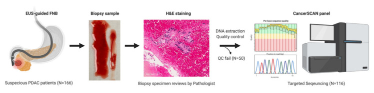
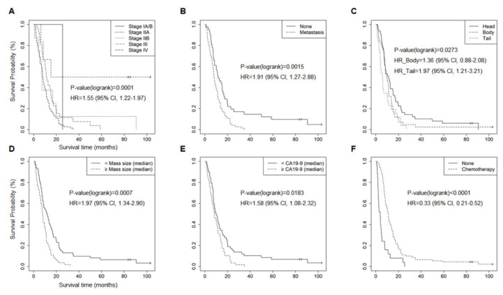
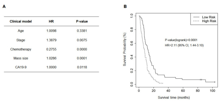
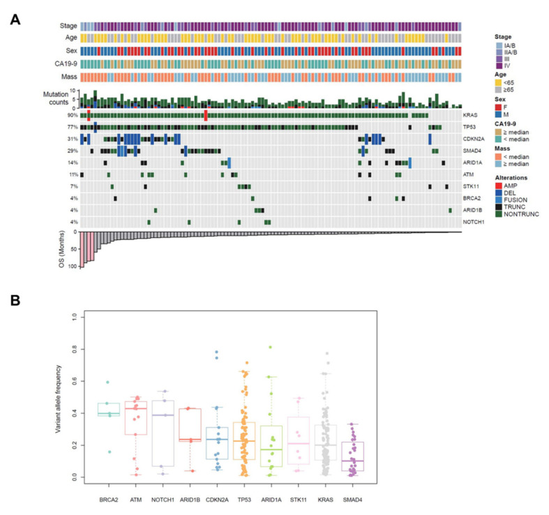
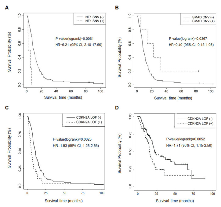
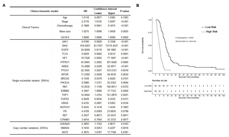
The aim of this study was to investigate the clinical utility of minimal specimens acquired from endoscopic ultrasound-guided fine-needle biopsy (EUS-FNB) and perform targeted deep sequencing as a prognosis prediction tool for pancreatic ductal adenocarcinoma (PDAC). A total of 116 specimens with pathologically confirmed PDAC via EUS-FNB were tested using CancerSCAN® panel for a customized targeted deep sequencing. Clinical prognostic factors significantly associated with survival in PDACs were as follows: stage, tumor mass size, tumor location, metastasis, chemotherapy, and initial CA19-9 level. A total of 114 patients (98.3%) had at least a single genetic alteration, and no mutations were detected in two patients, although they were qualified for the targeted deep sequencing. The frequencies of major gene mutations responsible for PDACs were KRAS 90%, CDKN2A 31%, TP53 77%, and SMAD4 29%. A somatic point mutation of NF1, copy number alteration of SMAD4, and loss-of-function of CDKN2A were significantly associated genetic factors for overall survival. Moreover, BRCA2 point mutation was related to liver metastasis. Finally, a clinico-genomic model was developed to estimate the prognosis of patients with PDAC based on clinical parameters and genetic alterations affecting survival in patients; 20 single nucleotide variants and three copy number variations were selected. Targeted deep sequencing on minimal specimens of PDACs was performed, and it was applied to establish a clinico-genomic model for prognosis prediction.

Keywords: clinico-genomic model; endoscopic ultrasound-guided fine needle core biopsy; pancreatic ductal adenocarcinoma; prognosis prediction; targeted deep sequencing.


Conflict of interest statement

There is no conflict to disclose.

Figures

Figure 1
Figure 1
The schematic flow of study design. Patients suspected of PDAC (N = 166) performed endoscopic ultrasound-guided fine-needle biopsy (EUS-FNB) to obtain pancreas specimens, which were examined by pathologists after hematoxylin & eosin (H&E) staining. DNAs were extracted from each PDAC specimen, and DNA analytes (N = 116) passing a quality control (QC) test were sequenced by using CancerSCAN® panel. PDAC, pancreatic ductal adenocarcinoma.
Figure 2
Figure 2
Kaplan–Meier plots for clinical factors significantly associated with survival. The Kaplan–Meier plots demonstrate six clinical factors with a significant (p < 0.05) association with the survival of patients with PDAC. (A) Stages, (B) metastasis, (C) tumor location, (D) tumor mass size, (E) CA19-9 level, and (F) chemotherapy. Hazard ratio (HR), confidence interval (CI), and p-value were obtained from univariate Cox proportional hazard test.
Figure 3
Figure 3
Clinical model. A clinical model considering various clinical parameters were developed to predict the survival of patients with pancreatic ductal adenocarcinoma (PDAC). (A) Selected clinical factors to establish the clinical model. (B) Kaplan–Meier plot for the high-risk group and the low-risk group of PDAC patients predicted by the clinical model (p = 0.0001).
Figure 4
Figure 4
Landscape of genomic alterations identified by targeted deep sequencing in EUS-FNB specimens of patients with PDAC. (A) Co-mutation plot displaying integrated genomic data for 116 EUS-FNB specimens displayed as columns, including somatic mutations and somatic copy number variations for significantly mutated genes listed at the right (Top 10). The percentage of PDAC samples with an alteration of any type is noted at the left. Stage, age, sex, CA19-9 level, tumor mass size, and the number of any genetic alterations per each sample is presented on the top. Overall survival (OS) of each patient is shown on the bottom. (B) Variant allele frequency (VAF) distributions are analyzed, and median values are indicated by the central bar in the box and whiskers plots. PDAC, pancreatic ductal adenocarcinoma; EUS-FNB, endoscopic ultrasound-guided fine-needle biopsy; AMP, copy number amplification; DEL, copy number deletion; FUSION, structure variation; TRUNC, truncating mutation; NONTRUNC, non-truncating mutation.
Figure 5
Figure 5
Kaplan–-Meier plots for genetic alterations significantly associated with survival. The Kaplan–Meier plots indicate genes significantly (p < 0.05) associated with the survival of patients with pancreatic ductal adenocarcinoma (PDAC). (A) Single nucleotide variant (SNV) of NF1 (p = 0.0061), (B) copy number variation (CNV) of SMAD4 (p = 0.0367), and (C) loss-of-function (LOF) mutation of CDKN2A (p = 0.0025) were related to survival in PDAC. (D) LOF mutation of CDKN2A and worse prognosis was verified by TCGA data.
Figure 6
Figure 6
Clinico-genomic model. A clinico-genomic model considering clinical parameters and genomic alterations was developed to predict patient survival. (A) Selected factors to establish the clinico-genomic model. (B) Kaplan–Meier plot for the high-risk group and the low-risk group of patients with pancreatic ductal adenocarcinoma (PDAC) predicted by the clinico-genomic model (p < 0.0001).

Similar articles

Cited by

References

    1. Siegel R.L., Miller K.D., Jemal A. Cancer statistics, 2018. Ca A Cancer J. Clin. 2018;68:7–30. doi: 10.3322/caac.21442. - DOI - PubMed
    1. Jung K.W., Won Y.J., Kong H.J., Oh C.M., Cho H., Lee D.H., Lee K.H. Cancer statistics in Korea: Incidence, mortality, survival, and prevalence in 2012. Cancer Res. Treat. Off. J. Korean Cancer Assoc. 2015;47:127–141. doi: 10.4143/crt.2015.060. - DOI - PMC - PubMed
    1. Hidalgo M. Pancreatic cancer. N. Engl. J. Med. 2010;362:1605–1617. doi: 10.1056/NEJMra0901557. - DOI - PubMed
    1. Adham M., Jaeck D., Le Borgne J., Oussoultzouglou E., Chenard-Neu M.P., Mosnier J.F., Scoazec J.Y., Mornex F., Partensky C. Long-term survival (5–20 years) after pancreatectomy for pancreatic ductal adenocarcinoma: A series of 30 patients collected from 3 institutions. Pancreas. 2008;37:352–357. doi: 10.1097/MPA.0b013e31818166d2. - DOI - PubMed
    1. Meldrum C., Doyle M.A., Tothill R.W. Next-generation sequencing for cancer diagnostics: A practical perspective. Clin. Biochem. Rev. 2011;32:177–195. - PMC - PubMed