Review
. 2021 Jun 19;9(6):694.
doi: 10.3390/biomedicines9060694.

Histone H2B Mutations in Cancer

Affiliations
Review

Histone H2B Mutations in Cancer

Yi Ching Esther Wan et al. Biomedicines. .

Abstract

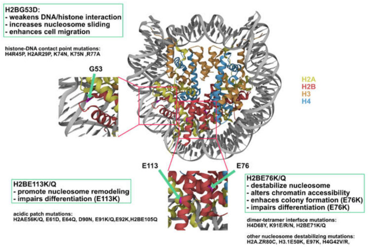
Oncohistones have emerged as a new area in cancer epigenetics research. Recent efforts to catalogue histone mutations in cancer patients have revealed thousands of histone mutations across different types of cancer. In contrast to previously identified oncohistones (H3K27M, H3G34V/R, and H3K36M), where the mutations occur on the tail domain and affect histone post-translational modifications, the majority of the newly identified mutations are located within the histone fold domain and affect gene expression via distinct mechanisms. The recent characterization of the selected H2B has revealed previously unappreciated roles of oncohistones in nucleosome stability, chromatin accessibility, and chromatin remodeling. This review summarizes recent advances in the study of H2B oncohistones and other emerging oncohistones occurring on other types of histones, particularly those occurring on the histone fold domain.

Keywords: H2B; cancer epigenetics; epigenetics; histone mutation; oncohistone.


Conflict of interest statement

The authors declare no conflict of interest.

Figures

Figure 1
Figure 1
Secondary structure of the four core histones. Histones generally consist of two disordered tails bracketing the histone fold domain and some additional structured fold unique to each type of histone. Helices are represented by rectangles and loops are represented by lines.
Figure 2
Figure 2
Schematics of immunoblots depicting the trans-inhibitory effect of H3K27M/H3K36M and cis-inhibitory effect of H3G34V/R. A tagged version of H3.3 (wildtype or mutated) was expressed in a human cell line followed by immunoprecipitation of the tagged H3.3 to retrieve mono-nucleosomes containing both endogenous H3.3 and tagged H3.3 (heterotypic nucleosome). For H3K27M and H3K36M (lane 2–3), the reduction of H3K27me3/H3K36me3 are observed on both copies of H3.3 (trans-inhibition). In contrast, the reduction of H3K36me3 is observed only on the tagged H3.3 (cis-inhibition). Of note, the expression of H3K36M also leads to an increase in H3K27me3 on endogenous H3.3. This figure is adapted from [10,15].
Figure 3
Figure 3
Illustration showing the H2A–H2B L1–L2 DNA binding region. H2AR77 (light blue) inserts into the minor groove of DNA. H2AT76 and I78, the two residues flanking H2AR77, form hydrogen bonds with H2BG53 (PDB code 2CV5).
Figure 4
Figure 4
Restriction enzyme accessibility assay evaluating chromatin remodeling activity. The chromatin remodeler remains inactive in the absence of ATP. The restriction enzyme cut site is hence protected by the octamer (upper panel). The addition of ATP activates the chromatin remodeler, resulting in nucleosome sliding and exposure of the cut site (lower panel).
Figure 5
Figure 5
Illustration highlighting the H2B–H4 interface. H2BE76 locates in the inner region of the nucleosome and interacts with H4R92 (PDB code 2CV5).
Figure 6
Figure 6
Left: Illustration showing the location of the acidic patch. The eight acidic patch residues are represented by bright yellow spheres. Right: Detailed view of acidic patch residues whose mutations have been shown to affect nucleosome sliding. Residues that inhibit nucleosome sliding when mutated are highlighted in orange. Residues that promote nucleosome sliding when mutated are highlighted in blue (PDB code: 2CV5).
Figure 7
Figure 7
Cartoon illustration of nucleosome symmetry and orientations.
Figure 8
Figure 8
Cartoon illustrating the result of chromatin remodeling assay conducted by Dao et al. [59]. Heterotypic nucleosomes of opposite orientations behave differently in SMARCA5-dependent chromatin remodeling assay. Hetero-anti-Nuc3A (mutant histone on the opposite side of DNA overhang) shows similar sliding as wildtype nucleosome, while hetero-syn-Nuc3A and homo- Nuc3A are completely inactive. H2A-3A, triple H2A mutant of E61A, D90A, E92A.
Figure 9
Figure 9
H2B mutations represent three novel classes of oncohistones. Nucleosome (PDB code 2CV5) showing the location of H2BG53, E76, and E113 (highlighted in magenta).

References

    1. Zhou K., Gaullier G., Luger K. Nucleosome structure and dynamics are coming of age. Nat. Struct. Mol. Biol. 2019;26:3–13. doi: 10.1038/s41594-018-0166-x. - DOI - PMC - PubMed
    1. Weinberg D.N., Allis C.D., Lu C. Oncogenic mechanisms of histone H3 mutations. Cold Spring Harb. Perspect. Med. 2017;7:a026443. doi: 10.1101/cshperspect.a026443. - DOI - PMC - PubMed
    1. Mohammad F., Helin K. Oncohistones: Drivers of pediatric cancers. Genes Dev. 2017;31:2313–2324. doi: 10.1101/gad.309013.117. - DOI - PMC - PubMed
    1. Schwartzentruber J., Korshunov A., Liu X.-Y., Jones D.T., Pfaff E., Jacob K., Sturm D., Fontebasso A.M., Quang D.-A.K., Tönjes M., et al. Driver mutations in histone H3. 3 and chromatin remodelling genes in paediatric glioblastoma. Nature. 2012;482:226–231. doi: 10.1038/nature10833. - DOI - PubMed
    1. Sturm D., Witt H., Hovestadt V., Khuong-Quang D.-A., Jones D.T., Konermann C., Pfaff E., Tönjes M., Sill M., Bender S., et al. Hotspot mutations in H3F3A and IDH1 define distinct epigenetic and biological subgroups of glioblastoma. Cancer Cell. 2012;22:425–437. doi: 10.1016/j.ccr.2012.08.024. - DOI - PubMed

LinkOut - more resources