. 2021 Jun 21;22(12):6621.
doi: 10.3390/ijms22126621.

Electrophysiology of hiPSC-Cardiomyocytes Co-Cultured with HEK Cells Expressing the Inward Rectifier Channel

Affiliations

Electrophysiology of hiPSC-Cardiomyocytes Co-Cultured with HEK Cells Expressing the Inward Rectifier Channel

Ana Da Silva Costa et al. Int J Mol Sci. .

Abstract

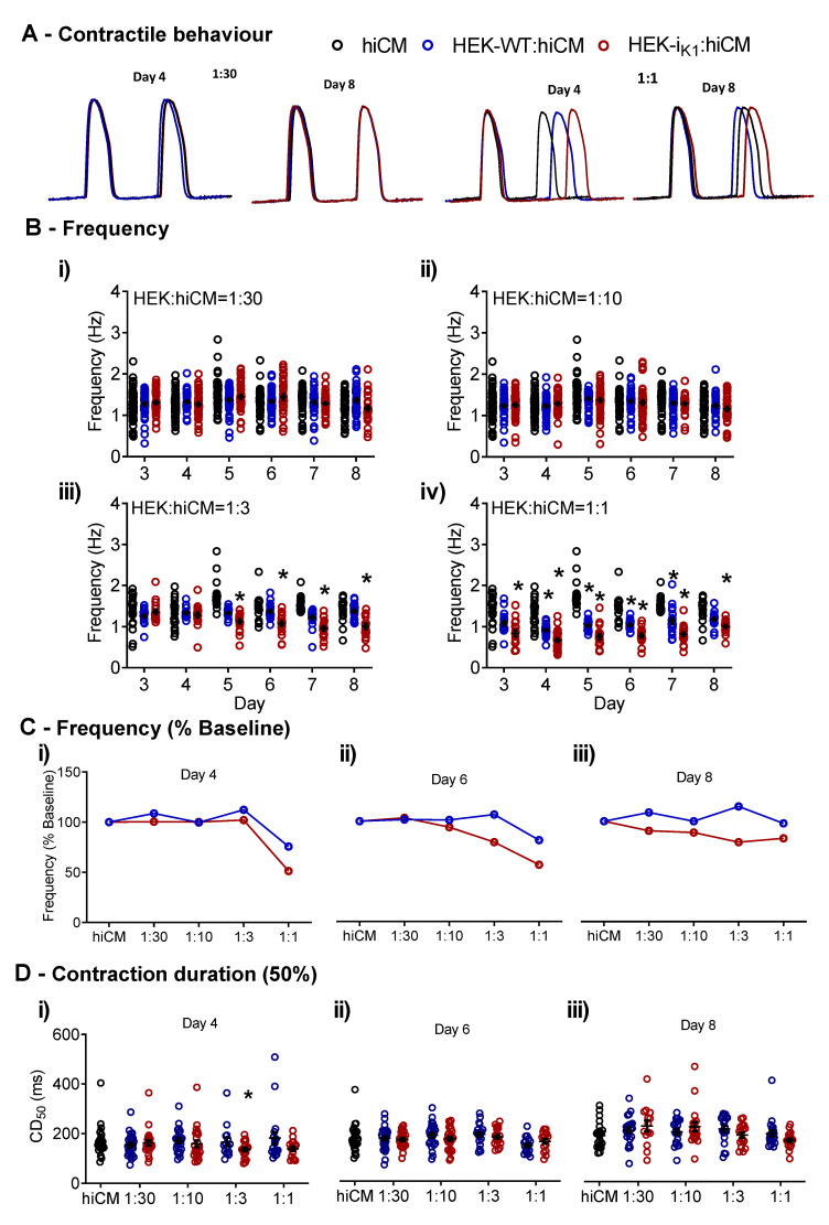
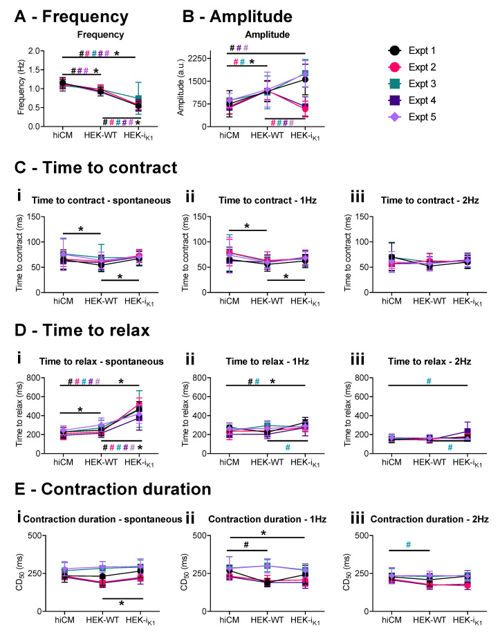
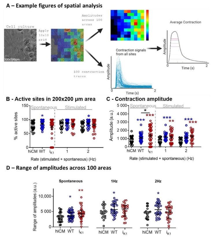
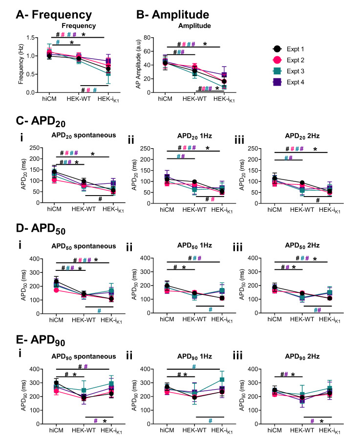
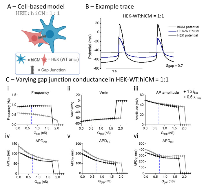
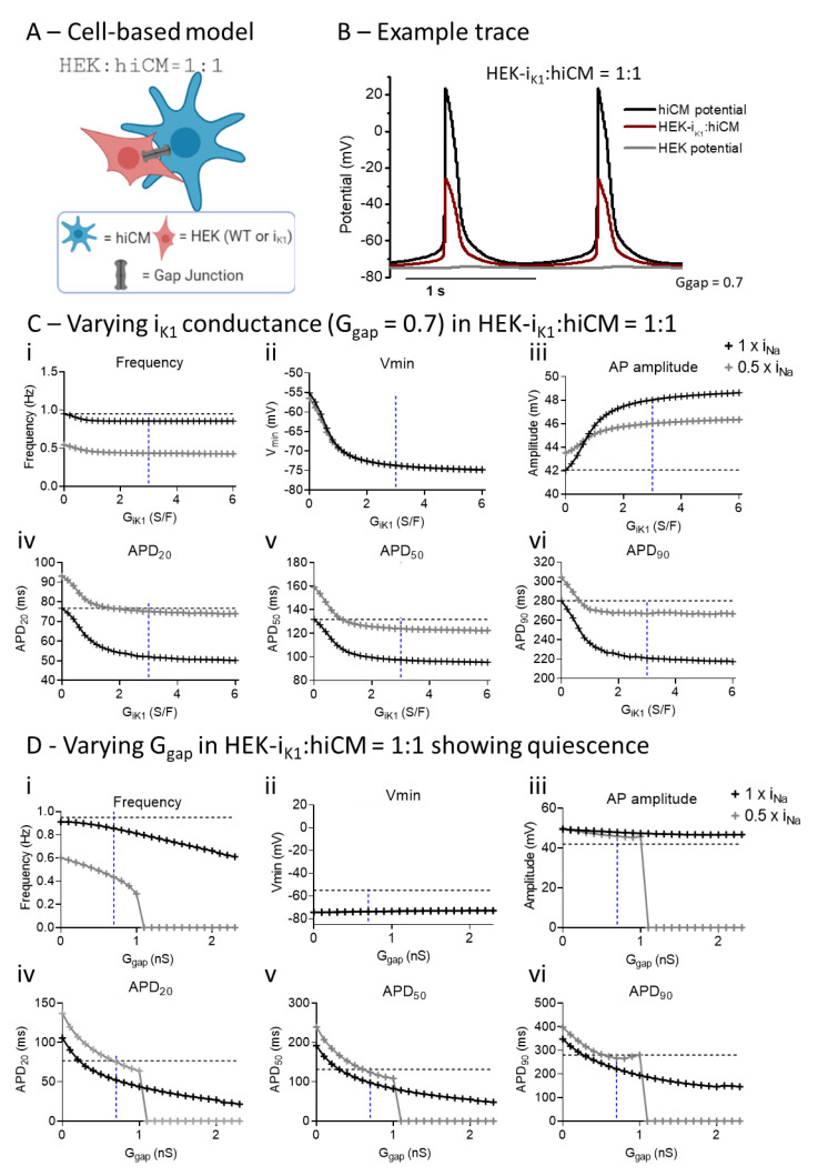
The immature electrophysiology of human-induced pluripotent stem cell-derived cardiomyocytes (hiCMs) complicates their use for therapeutic and pharmacological purposes. An insufficient inward rectifying current (IK1) and the presence of a funny current (if) cause spontaneous electrical activity. This study tests the hypothesis that the co-culturing of hiCMs with a human embryonic kidney (HEK) cell-line expressing the Kir2.1 channel (HEK-IK1) can generate an electrical syncytium with an adult-like cardiac electrophysiology. The mechanical activity of co-cultures using different HEK-IK1:hiCM ratios was compared with co-cultures using wildtype (HEK-WT:hiCM) or hiCM alone on days 3-8 after plating. Only ratios of 1:3 and 1:1 showed a significant reduction in spontaneous rate at days 4 and 6, suggesting that IK1 was influencing the electrophysiology. Detailed analysis at day 4 revealed an increased incidence of quiescent wells or sub-areas. Electrical activity showed a decreased action potential duration (APD) at 20% and 50%, but not at 90%, alongside a reduced amplitude of the aggregate AP signal. A computational model of the 1:1 co-culture replicates the electrophysiological effects of HEK-WT. The addition of the IK1 conductance reduced the spontaneous rate and APD20, 50 and 90, and minor variation in the intercellular conductance caused quiescence. In conclusion, a 1:1 co-culture HEK-IK1:hiCM caused changes in electrophysiology and spontaneous activity consistent with the integration of IK1 into the electrical syncytium. However, the additional electrical effects of the HEK cell at 1:1 increased the possibility of electrical quiescence before sufficient IK1 was integrated into the syncytium.

Keywords: HEK; IK1; co-culture; electrophysiology; hiCMs; maturation.


Conflict of interest statement

The funders had no role in the design of the study; in the collection, analyses, or interpretation of data; in the writing of the manuscript, or in the decision to publish the results.

Figures

Figure 1
Figure 1
The effects of co-cultures of hiCMs with increasing densities of HEK, for both HEK-IK1:hiCM and HEK–WT:hiCM. (A) Example traces of the effects of 1:30 and 1:1 at days 4 and 8. (Bi) Frequency of spontaneous contraction at days 4 to 8 with the ratio 1:30; (Bii) frequency at the 1:10 ratio; (Biii) frequency at the 1:3 ratio; (Biv) frequency at the 1:1 ratio; (C) frequency as a percentage from baseline (standard hiCM culture) for the range of ratios. (Ci) Day 4; (Cii) day 6; (Ciii) day 8. (D) Contraction duration (CD50) on days 4, 6 and 8 comparing hiCM culture, HEK–WT:hiCM and HEK-IK1:hiCM. One-way ANOVA with Bartlett’s test, * p < 0.05, n = 51 wells.
Figure 2
Figure 2
Cell movement effects of the 1:1 co-culture on day 4 in vitro in serum-free conditions, showing observations from individual experiments. Each plating is shown in a different color, and data are presented as mean+/−SD. (A) Frequency of spontaneous contraction. (B) Amplitude of the spontaneous contraction. (C) Time to contract at (Ci) spontaneous, (Cii) 1 Hz, and (Ciii) 2 Hz conditions. (D) Time to relax at (Di) spontaneous, (Dii) 1 Hz, and (Diii) 2 Hz. (E) This shows 50% of the contraction duration at (Ei) spontaneous, (Eii) 1 Hz, and (Eiii) 2 Hz. The comparison of each culture (hiCM, HEK–WT:hiCM and HEK-IK1:hiCM) within each experiment is shown as # in the respective experimental, and 2-way ANOVA was used for statistical analysis. The mean of all experiments was then compared using a paired t-test and this is shown as * (p < 0.05).
Figure 3
Figure 3
Spatial analysis of 1:1 HEK:CM on day 4 in culture in serum-free medium. (A) Example images obtained using spatial analysis of contractile motion. A heat map showing amplitudes recorded in a 200 × 200 µm area using a 10 × 10 grid were derived from the brightfield image (left). The colors represent the amplitude of the contraction in each square. Contraction signals from all sites and the average contraction are also shown. (B) Percentage of active sites representing contractile cells in a 200 × 200 µm area. (C) The amplitude of the contraction determined by pixel displacement. (D) Mean of range of amplitudes across a 10 × 10 grid. Unpaired t-test: HEK-IK1 vs. HEK–WT (black) and hiCM vs. HEK–WT (blue) or HEK-IK1 (red). *** p < 0.001, ** p < 0.01, * p < 0.05, n > 28 cells, 5 platings.
Figure 4
Figure 4
Electrophysiological effects of 1:1 co-culture on day 4 in vitro in serum-free conditions, showing the observations from individual experiments. Each plating is shown in a different color, and data are presented as mean+/−SD. (A) Frequency of spontaneous beating. (B) Amplitude of the AP. (C) APD20 at (Ci) spontaneous, (Cii) 1Hz, and (Ciii) 2Hz. (D) APD50 at (Di) spontaneous, (Dii) 1 Hz, and (Diii) 2 Hz. (E) APD90 at (Ei) spontaneous, (Eii) 1 Hz, and (Eiii) 2 Hz. Due to the small AP amplitude and low signal-to-noise ratio (SNR) (Supplementary Figure S3) in HEK-IK1:hiCM, fewer data are available for APD90 at spontaneous rates. Comparison of each culture (hiCM, HEK–WT:hiCM and HEK-IK1:hiCM) within each experiment is shown as # in the respective experimental color, and 2-way ANOVA was used for statistical analysis. The mean of all experiments was then compared using a paired t-test and this is shown with * (p < 0.05).
Figure 5
Figure 5
Computational modeling shows the effects of varying gap junction conductance (Ggap) in a co-culture of hiCM with HEK–WT, and different Na+ channel expression. (A) Diagram of HEK:hiCM interaction used for computational model. (B) Example trace showing hiCM potential, HEK potential, and the average of the two. (C) Varying Ggap effects on frequency of spontaneous beating, hiCM’s Vmin, amplitude and APD. Simulations were done in regular INa (1 × INa) and when only half (0.5 × INa) was present.
Figure 6
Figure 6
Computational modeling shows the effects of varying gap junction conductance (Ggap) and IK1 conductance (GIK1) in co-cultures of hiCM with HEK-IK1 and different Na+ channel expressions. (A) Diagram of HEK:hiCM interaction used for computational model. (B) Example trace showing hiCM potential, HEK potential, and the average of the two. (C) Different parameters are affected by varying IK1 conductance in a full INa model, and a half INa. (D) Electrophysiology is affected by increasing gap junction conductance in a co-culture of hiCM with HEK-IK1.

References

    1. Verkerk A.O., Veerman C.C., Zegers J.G., Mengarelli I., Bezzina C.R., Wilders R. Patch-clamp recording from human induced pluripotent stem cell-derived cardiomyocytes: Improving action potential characteristics through dynamic clamp. Int. J. Mol. Sci. 2017;18:1873. doi: 10.3390/ijms18091873. - DOI - PMC - PubMed
    1. Casini S., Verkerk A.O., Remme C.A. Human iPSC-derived cardiomyocytes for investigation of disease mechanisms and therapeutic strategies in inherited arrhythmia syndromes: Strengths and limitations. Cardiovasc. Drugs Ther. 2017;31:325–344. doi: 10.1007/s10557-017-6735-0. - DOI - PMC - PubMed
    1. Hoekstra M., Mummery C.L., Wilde A.A.M., Bezzina C.R.P., Verkerk A.O. Induced pluripotent stem cell derived cardiomyocytes as models for cardiac arrhythmias. Front. Physiol. 2012;3:346. doi: 10.3389/fphys.2012.00346. - DOI - PMC - PubMed
    1. Burridge P.W., Keller G., Gold J.D., Wu J.C. Production of de novo cardiomyocytes: Human pluripotent stem cell differentiation and direct reprogramming. Cell Stem Cell. 2012;10:16–28. doi: 10.1016/j.stem.2011.12.013. - DOI - PMC - PubMed
    1. Matsa E., Burridge P.W., Wu J.C. Human stem cells for modeling heart disease and for drug discovery. Sci. Transl. Med. 2014;6:239ps6. doi: 10.1126/scitranslmed.3008921. - DOI - PMC - PubMed

Publication types

Substances

LinkOut - more resources