. 2021 Jun 21;10(6):1563.
doi: 10.3390/cells10061563.

Advanced Maternal Age Deteriorates the Developmental Competence of Vitrified Oocytes in Mice

Affiliations

Advanced Maternal Age Deteriorates the Developmental Competence of Vitrified Oocytes in Mice

Ju Hee Lee et al. Cells. .

Abstract

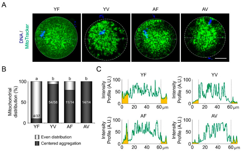
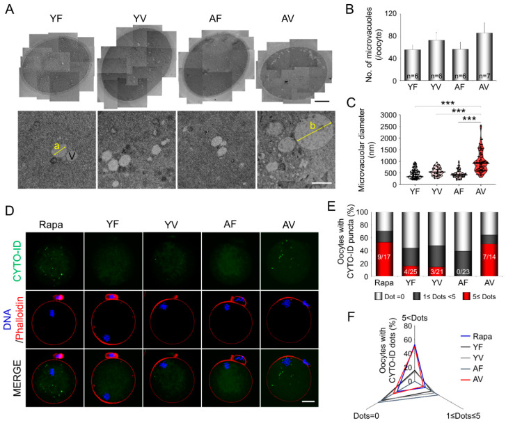
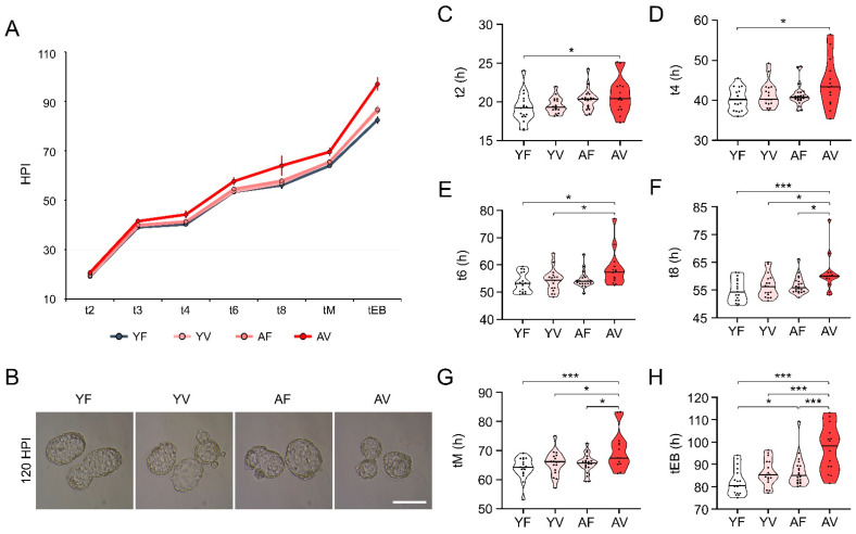
Advanced maternal age (AMA) is known to be related to the decrease in the quality and quantity of oocytes. Oocyte vitrification is now considered an established assisted reproductive technology for fertility preservation. However, it remains unclear whether the oocytes in older women are more sensitive to various insults during vitrification. Thus, we evaluated whether AMA affects cellular and molecular features and developmental outcomes of oocytes after vitrification in mice. The oocytes were grouped as young fresh (YF), young vitrified/warmed (YV), aged fresh (AF), and aged vitrified/warmed (AV). The survival rate of AV oocytes was significantly lower than that of YV oocytes. The rates of fertilization, cleavage, and blastocyst formation of AV oocytes were significantly lower than those of other groups. AV oocytes were represented as aberrations in mitochondria distribution, microvacuole size, and autophagosome formation, leading to delayed embryo development in mice. This delay was associated with a reduced number of total cells and trophectoderm in the blastocyst developed from AV oocytes. Collectively, AMA exaggerates the vulnerability of oocytes to cryo-damage that occurs during vitrification in mice, suggesting that the current vitrification protocols optimized for oocytes from young females should be modified for oocytes from aged women.

Keywords: advanced maternal age (AMA); developmental competence; fertility preservation; oocyte quality; oocyte vitrification; time-lapse monitoring.


Conflict of interest statement

The authors declare no conflict of interest.

Figures

Figure 1
Figure 1
Age-dependent changes in the number and morphological abnormalities of ovulated oocytes. (A) A schematic comparison of the life span between humans and C57BL/6 (BL6) mice, which has been modified from “Mouse models in aging research” [23]. (B) The total number of ovulated oocytes from BL6 mice by age (from 1- to 18-month-old). The graph presents the average number of ovulated oocytes per mouse (n = 6–44 mice in each group). (C) An age-dependent increase in morphologically abnormal oocytes (1- to 18-month-old). White and black bars represent the percentage of normal and abnormal oocytes, respectively (n = 38–635 oocytes in each group). Red dots represent the median values in each group. (D) Representative microscopic images of ovulated oocytes from young and aged BL6 mice after superovulation. Red arrowheads indicate morphologically abnormal oocytes. (E) A schematic diagram to illustrate the experimental procedures to evaluate whether the survival rates of oocytes from young and aged mice are different between strains after vitrification. (F) Comparison of the survival rates of VW oocytes from young (1- to 2-month-old) and aged (10- to 12-month-old) mice. The graphs present the survival rate of total ovulated VW oocytes (n = 125–327 oocytes in each group). Scale bar = 75 µm. Statistical comparisons were performed using chi-squared test. *** p < 0.005.
Figure 2
Figure 2
Abnormal spindle and chromosome organization in oocytes from aged mice. (A) Representative images of immunofluorescence staining for α-tubulin (a spindle marker, Green), pericentrin (PCNT; a spindle pole marker, Red), and DAPI (DNA, Blue) in oocytes from young and aged mice with or without vitrification. The bottom panels show the enlarged images of the boxed areas in the upper panels. Arrowhead and arrow indicate abnormal spindle/spindle pole and chromosome, respectively. Note that abnormal spindle and chromosome organization were often observed in oocytes from aged mice. (B) Quantitative analyses of oocytes with abnormal spindle and chromosomal morphologies (n = 10–23 oocytes in each group). White and black bars represent the percentage of oocytes with normal and abnormal morphologies, respectively Scale bar = 20 µm. Statistical analyses were performed using Fisher’s exact test. * p < 0.05. YF: Young fresh, YV: Young vitrified, AF: Aged fresh, AV: Aged vitrified.
Figure 3
Figure 3
Fluorescence live cell imaging to examine the patterns of mitochondrial distribution after vitrification in oocytes. (A) Representative microscopic images of mitochondrial distribution visualized using MitoTracker dye (Green) in live oocytes from young and aged mice before or after vitrification. (B) Quantitative analyses of mitochondrial distribution patterns (n = 14–58 oocytes in each group). Mitochondrial distribution was divided into even distribution and centered aggregation. White and black bars represent the percentage of even distribution and centered aggregation, respectively. (C) The graphs present the mitochondrial intensity profiles throughout the ooplasm of oocytes. Scale bar = 20 µm. Statistical analyses were performed using Fisher’s exact test. Different superscript letters (a–b) denote a significant difference from the control, p < 0.05. YF: Young fresh, YV: Young vitrified, AF: Aged fresh, AV: Aged vitrified.
Figure 4
Figure 4
Ultrastructural analyses of fresh and vitrified/warmed (VW) oocytes from young and aged mice. (A) Transmission electron microscopy images showing the microvacuoles in fresh and VW oocytes (a = 722 nm, b = 1910 nm). Upper panel scale bar = 12 µm; Bottom panel scale bar = 1 µm. (B,C) Quantitative analyses for the diameter and number of microvacuoles in fresh and VW oocytes from young and aged mice (n = 6–7 oocytes in each group). Note that the size and number of microvacuoles increased in VW oocytes from aged mice. The horizontal black lines represent the median values of the microvacuolar diameter of oocytes. Statistical comparisons were performed using one-way ANOVA, followed by Tukey’s multiple comparisons test and Dunn’s multiple comparisons test. V: Microvacuole. *** p < 0.005. (D) Representative microscopic images of autophagosome visualized using CYTO-ID (Green) in oocytes from young and aged mice before or after vitrification. Confocal imaging analysis of the oocytes. (E,F) Quantitative analysis of CYTO-ID puncta in fresh and VW oocytes from young and aged mice (n = 14–25 oocytes in each group). YF: Young fresh, YV: Young vitrified, AF: Aged fresh, AV: Aged vitrified.
Figure 5
Figure 5
Effects of vitrification on the development of embryos from in vitro fertilization of oocytes from young and aged mice. (AC) The rates of fertilization, cleavage, and blastocyst formation of oocytes from young (n = 10 mice, 1- to 2-month-old) and aged (n = 30 mice, 10- to 12-month-old) mice (n = 64–105 oocytes in each group) after in vitro fertilization. Statistical comparisons were performed using chi-squared test. ** p < 0.01, *** p < 0.005. YF: Young fresh, YV: Young vitrified, AF: Aged fresh, AV: Aged vitrified.
Figure 6
Figure 6
Comparison of embryo morphokinetics of fresh and vitrified/warmed (VW) oocytes from young and aged mice. (AH) Comparison of morphokinetics of embryos developed from oocytes from young and aged mice after vitrification (n = 14–20 oocytes in each group). t2, t3, t4, t6, t8: time in hours post insemination (HPI) for the embryo to reach the 2-, 3-, 4-, 6-, 8-cell stage, respectively, tM: time for compaction, tEB: time for the embryo to start blastocoel formation. Statistical comparisons were performed using student’s t-test. Four independent experiments were performed with similar results. * p < 0.05, *** p < 0.005. Scale bar = 140 µm. YF: Young fresh, YV: Young vitrified, AF: Aged fresh, AV: Aged vitrified.
Figure 7
Figure 7
Evaluation of the effects of vitrification on blastocyst development in oocytes after in vitro fertilization. (A) Immunofluorescence staining for OCT4 [inner cell mass (ICM) marker], and DAPI (nuclei) in normal and abnormal blastocysts developed from fresh and vitrified/warmed oocytes from young and/or aged mice (n = 13–40 blastocysts in each group). (BE) Quantitative analyses of the total cell number, trophectoderm (TE), ICM, and ICM/TE ratio in blastocysts from vitrified/warmed oocytes of young and aged mice. The total cell number was calculated by counting the DAPI-stained nuclei. The number of ICM and TE were counted from OCT4-positive and DAPI-positive cells without OCT4, respectively. The horizontal black lines represent the median values of total cell number, number of ICM and TE, and ICM/TE ratio. Statistical comparisons were performed using one-way ANOVA, followed by Tukey’s multiple comparisons test. Scale bar = 50 µm. * p < 0.05. YF: Young fresh, YV: Young vitrified, AF: Aged fresh, AV: Aged vitrified.

Similar articles

Cited by

References

    1. Cobo A., García-Velasco J.A., Remohí J., Pellicer A. Oocyte vitrification for fertility preservation for both medical and nonmedical reasons. Fertil. Steril. 2021;115:1091–1101. doi: 10.1016/j.fertnstert.2021.02.006. - DOI - PubMed
    1. Pantazis A., Clark S.J. A parsimonious characterization of change in global age-specific and total fertility rates. PLoS ONE. 2018;13:e0190574. doi: 10.1371/journal.pone.0190574. - DOI - PMC - PubMed
    1. Ubaldi F.M., Cimadomo D., Vaiarelli A., Fabozzi G., Venturella R., Maggiulli R., Mazzilli R., Ferrero S., Palagiano A., Rienzi L. Advanced maternal age in IVF: Still a challenge? The present and the future of its treatment. Front. Endocrinol. 2019;10:94. doi: 10.3389/fendo.2019.00094. - DOI - PMC - PubMed
    1. Cimadomo D., Fabozzi G., Vaiarelli A., Ubaldi N., Ubaldi F.M., Rienzi L. Impact of maternal age on oocyte and embryo competence. Front. Endocrinol. 2018;9:327. doi: 10.3389/fendo.2018.00327. - DOI - PMC - PubMed
    1. Johnston M., Richings N.M., Leung A., Sakkas D., Catt S. A major increase in oocyte cryopreservation cycles in the USA, Australia and New Zealand since 2010 is highlighted by younger women but a need for standardized data collection. Hum. Reprod. 2020;36:624–635. doi: 10.1093/humrep/deaa320. - DOI - PubMed

Publication types

LinkOut - more resources