. 2021 Jun 1;10(6):1362.
doi: 10.3390/cells10061362.

Pulmonary Fibroelastotic Remodelling Revisited

Affiliations

Pulmonary Fibroelastotic Remodelling Revisited

Peter Braubach et al. Cells. .

Abstract

Pulmonary fibroelastotic remodelling occurs within a broad spectrum of diseases with vastly divergent outcomes. So far, no comprehensive terminology has been established to adequately address and distinguish histomorphological and clinical entities. We aimed to describe the range of fibroelastotic changes and define stringent histological criteria. Furthermore, we wanted to clarify the corresponding terminology in order to distinguish clinically relevant variants of pulmonary fibroelastotic remodelling. We revisited pulmonary specimens with fibroelastotic remodelling sampled during the last ten years at a large European lung transplant centre. Consensus-based definitions of specific variants of fibroelastotic changes were developed on the basis of well-defined cases and applied. Systematic evaluation was performed in a steps-wise algorithm, first identifying the fulcrum of the respective lesions, and then assessing the morphological changes, their distribution and the features of the adjacent parenchyma. We defined typical alveolar fibro-elastosis as collagenous effacement of the alveolar spaces with accompanying hyper-elastosis of the remodelled and paucicellular alveolar walls, independent of the underlying disease in 45 cases. Clinically, this pattern could be seen in (idiopathic) pleuroparenchymal fibro-elastosis, interstitial lung disease with concomitant alveolar fibro-elastosis, following hematopoietic stem cell and lung transplantation, autoimmune disease, radio-/chemotherapy, and pulmonary apical caps. Novel in-transit and activity stages of fibroelastotic remodelling were identified. For the first time, we present a comprehensive definition of fibroelastotic remodelling, its anatomic distribution, and clinical associations, thereby providing a basis for stringent patient stratification and prediction of outcome.

Keywords: alveolar fibroelastosis; interstitial fibrosis; lung.


Conflict of interest statement

The authors declare no conflict of interest.

Figures
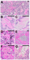
Figure 1
Figure 1
Typical histological patterns of alveolar fibroelastosis (AFE). (A) Typical AFE is characterized by a complete obliteration of the alveolar lumen with collagenous material with formation of either coarse (B) or fine (C) fibrils. In some cases, aggregates of macrophages containing anthracotic pigment can be observed (D). Lymphoid aggregates are a common finding in or at the border of AFE lesions (E). Increased cellularity with presence of mesenchymal cells (F) or lymphocytes (G) can be observed in the fibrotic areas to a variable degree. All images are elastic van Gieson stainings. Scale bars are 100 µm each.
Figure 2
Figure 2
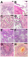
Typical histological patterns in special association with alveolar fibroelastosis (AFE). A set of typical features regularly found in spatial association with AFE: (A) Pronounced fibrosis of the visceral pleura. (B) Emphysema with an irreversible loss of alveolar septa. (C) Elastosis of the alveolar wall with incomplete alveolar fibrosis of the alveolar lumen with residual airspaces lined by cuboidal epithelium (*). (D) Fibroelastic interstitial expansion of the alveolar septa adjacent to the AFE lesion. (E) Aggregates of intraalveolar macrophages. (F) Cholesterol granulomas with multinucleated giant cells with clefts of cholesterol crystals. (G) Bronchiolitis obliterans with fibrous obliteration of small airways and (H) sclerosis of pulmonary arteries with hypertrophy of the media and intimal hyperplasia. Images are elastic van Gieson stains. Scale bars are 100 µm each.
Figure 3
Figure 3
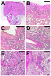
Compartmental distribution of alveolar fibroelastosis (AFE). AFE is commonly found in the subpleural parenchyma (A,B), in parabronchial (C) and paravascular (D) distribution and along interlobular septa (*, E). When not in association with these structures, we classified the localization as centrolobular (F). Images are haematoxylin and eosin (A) and elastic van Gieson (BF) stains. Scale bars are 500 µm each.
Figure 4
Figure 4
Radiological correlate of AFE Illustration of the two typical radiological patterns accompanying a histologic AFE diagnosis. (A). Radiological PPFE pattern with typical (sub)pleural distribution of fibrosis. (B) Apical cap.
Figure 5
Figure 5
Patterns of compartmental distribution of alveolar fibroelastosis (AFE): AFE is a pattern defined by the typical fibrous obliteration of the alveolar airspace with hyper-elastosis of the preserved alveolar structure. Similar histologic patterns can be observed in a variety of diseases which are distinguishable by a characteristic distribution of the AFE pattern. When AFE is found circumscript in the subpleural parenchyma of the upper lobe without any other indication of interstitial lung disease (ILD), a prognostic favourable pulmonal apical cap (PAC) is the most likely diagnosis. Further, circumscript focal AFE can be found e.g., after radiotherapy and around (unspecific) scars of the lung parenchyma. When found in association with other ILD patterns (e.g., usual interstitial pneumonia, UIP) the prognosis of the patient may be worse than when concomitant AFE is not found. AFE as dominant pattern with subpleural, parabronchial and paraseptal distribution and accentuation in the upper compartments of the lung is indicative of pleuroparenchymal fibroelastosis (PPFE), a rare ILD with poor prognosis.

References

    1. Parra E.R., Kairalla R.A., De Carvalho C.R.R., Capelozzi V.L. Abnormal deposition of collagen/elastic vascular fibres and prognostic significance in idiopathic interstitial pneumonias. Thorax. 2007;62:428–437. doi: 10.1136/thx.2006.062687. - DOI - PMC - PubMed
    1. Wynn T.A., Ramalingam T.R. Mechanisms of fibrosis: Therapeutic translation for fibrotic disease. Nat. Med. Nat. Publ. Group. 2012;18:1028–1040. doi: 10.1038/nm.2807. - DOI - PMC - PubMed
    1. Rosenbaum J.N., Butt Y.M., Johnson K.A., Meyer K., Batra K., Kanne J.P., Torrealba J.R. Pleuroparenchymal fibroelastosis: A pattern of chronic lung injury. Hum. Pathol. 2015;46:137–146. doi: 10.1016/j.humpath.2014.10.007. - DOI - PubMed
    1. Frankel S.K., Cool C.D., Lynch D.A., Brown K.K. Idiopathic Pleuroparenchymal Fibroelastosis. Chest. 2004;126:2007–2013. doi: 10.1378/chest.126.6.2007. - DOI - PubMed
    1. Travis W.D., Costabel U., Hansell D.M., King T.E., Lynch D.A., Nicholson A.G., Ryerson C.J., Ryu J.H., Selman M., Wells A.U., et al. An Official American Thoracic Society/European Respiratory Society Statement: Update of the International Multidisciplinary Classification of the Idiopathic Interstitial Pneumonias. Am. J. Respir. Crit. Care Med. 2013;188:733–748. doi: 10.1164/rccm.201308-1483ST. - DOI - PMC - PubMed

Publication types

MeSH terms