. 2021 Jun 1;13(6):822.
doi: 10.3390/pharmaceutics13060822.

Nanoparticle-Mediated Angiotensin-(1-9) Drug Delivery for the Treatment of Cardiac Hypertrophy

Affiliations

Nanoparticle-Mediated Angiotensin-(1-9) Drug Delivery for the Treatment of Cardiac Hypertrophy

Sabrina Sepúlveda-Rivas et al. Pharmaceutics. .

Abstract

Ang-(1-9) peptide is a bioactive vasodilator peptide that prevents cardiomyocyte hypertrophy in vitro and in vivo as well as lowers blood pressure and pathological cardiovascular remodeling; however, it has a reduced half-life in circulation, requiring a suitable carrier for its delivery. In this work, hybrid nanoparticles composed of polymeric nanoparticles (pNPs) based on Eudragit® E/Alginate (EE/Alg), and gold nanospheres (AuNS), were developed to evaluate their encapsulation capacity and release of Ang-(1-9) under different experimental conditions. Hybrid pNPs were characterized by dynamic light scattering, zeta potential, transmission and scanning electron microscopy, size distribution, and concentration by nanoparticle tracking analysis. Nanometric pNPs, with good polydispersity index and colloidally stable, produced high association efficiency of Ang-(1-9) and controlled release. Finally, the treatment of neonatal cardiomyocytes in culture with EE/Alg/AuNS 2% + Ang-(1-9) 20% pNPs decreased the area and perimeter, demonstrating efficacy in preventing norepinephrine-induced cardiomyocyte hypertrophy. On the other hand, the incorporation of AuNS did not cause negative effects either on the cytotoxicity or on the association capacity of Ang-(1-9), suggesting that the hybrid carrier EE/Alg/AuNS pNPs could be used for the delivery of Ang-(1-9) in the treatment of cardiovascular hypertrophy.

Keywords: angiotensin-(1-9); cardiac hypertrophy; gold nanoparticles; peptide delivery; polymeric nanoparticles.


Conflict of interest statement

The authors declare no conflict of interest.

Figures

Figure 1
Figure 1
SEM images of EE/Alg (CR = 0.5; TC = 15) pNPs obtained at 30,000× and 60,000×, bar represents 4 μm and 2 μm, respectively.
Figure 2
Figure 2
Transmission electron microscopy images of EE/Alg pNPs loaded with AuNS (TC = 15: CR = 1). Bars represent 200 nm (left) and 400 nm (right).
Figure 3
Figure 3
Graphs of (a) size and (b) concentration of empty EE/Alg pNPs and 2% EE/Alg/AuNS hybrid pNPs + 20% Ang-(1-9). The data represent the mean ± SD (n = 3), post-student t-test ns = not statistically significant with respect to empty pNPs.
Figure 4
Figure 4
Efficiency of association of (a) Ang-(1-9) 20% in EE/Alg pNPs at different CR; (b) Ang-(1-9) in 2% EE/Alg/AuNS pNPs. The results were expressed in % EA, which is represented as mean ± SD (n = 3).
Figure 5
Figure 5
Stability parameters of the hybrid (a) mean diameter; (b) zeta potential; and (c) polydispersity index of pNPs at 25 °C and 37 °C in media (phosphate buffer pH 7.4 (PBS), DMEM supplemented with 10% FBS and plasma and at 0, 15, and 60 min. Milli-Q water was used as a control. The values correspond to the mean DS (n = 3). Two-way ANOVA, Bonferroni post-test, * p < 0.05 v/s nanohybrids in Milli-Q water at 0 min, # p < 0.05 nanohybrid in Milli-Q water at 15 min, & p < 0.05 nanohybrids in Milli-Q water at 60 min.
Figure 5
Figure 5
Stability parameters of the hybrid (a) mean diameter; (b) zeta potential; and (c) polydispersity index of pNPs at 25 °C and 37 °C in media (phosphate buffer pH 7.4 (PBS), DMEM supplemented with 10% FBS and plasma and at 0, 15, and 60 min. Milli-Q water was used as a control. The values correspond to the mean DS (n = 3). Two-way ANOVA, Bonferroni post-test, * p < 0.05 v/s nanohybrids in Milli-Q water at 0 min, # p < 0.05 nanohybrid in Milli-Q water at 15 min, & p < 0.05 nanohybrids in Milli-Q water at 60 min.
Figure 6
Figure 6
Release profile of non-encapsulated Ang-(1-9) and Ang-(1-9) from pNPs EE/Alg CR: 0.5 and 10, in a dialysis system suspended in PBS pH 7.4 and 37 °C in time intervals. Values are expressed as mean ± SD (n = 3).
Figure 7
Figure 7
Release profile of Ang-(1-9) from EE/Alg/AuNS 2 and 20% pNPs CR: 0.5, in a dialysis system suspended in PBS pH 7.4 and 37 °C at intervals of time. Values are expressed as mean ± SD (n = 3).
Figure 8
Figure 8
Effect on the area (a) and perimeter (b) of cardiomyocytes treatment with NE, Ang-(1-9), and hybrid pNPs. Ctrl 24 h: Control cardiomyocytes in their maintenance medium for 24 h, NE: cardiomyocytes + subsequent NE 10 µM, Ang-(1-9): cardiomyocytes + subsequent Ang-(1-9) 10 µM; Blank pNPs: cardiomyocytes + subsequent EE/Alg pNPs, Hybrid pNPs: cardiomyocytes + EE/Alg/AuNS/Ang-(1-9) pNPs. Data represent mean values ± SD (n = 3). An ANOVA–Dunnett was performed to compare the 24 h control with the different control treatments, PRISMA Graph * (p < 0.05), ** (p < 0.001).
Figure 9
Figure 9
Effect on the cardiomyocyte area (a) and perimeter (b) of the pre-incubation of different stimuli Ctrl 24 h: cardiomyocyte control in MM (maintenance medium) 24 h; NE: cardiomyocytes 24h + norepinephrine 10 μM; NE + Ang-(1-9): norepinephrine 10 μM + angiotensin-(1-9) 10 μM; NE + Blank pNPs: norepinephrine 10 μM + EE/Alg/Ang-(1-9) pNPs; NE + Hybrid pNPs: norepinephrine 10 μM + pNPs EE/Alg/AuNS/Ang-(1-9)/; Ang-(1-9): cardiomyocytes 24 h + Ang-(1-9) 10 μM. Data represent mean values ± SD (n = 3). An ANOVA–Dunnett was performed to compare the 24 h control with the different control treatments, PRISMA Graph * (p < 0.05), ** (p < 0.001).
Figure 10
Figure 10
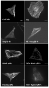
Images of the morphological changes that occur in the neonatal rat cardiomyocyte subjected to the different stimuli. Ctrl 24 h: cardiomyocyte control in MM (maintenance medium) 24 h; NE: cardiomyocytes 24 h + norepinephrine 10 μM; Ang-(1-9): cardiomyocytes 24h + Ang-(1-9) 10 μM; NE + Ang-(1-9): norepinephrine 10 μM + angiotensin-(1-9) 10 μM; NE + Blank pNPs: norepinephrine 10 μM + EE/Alg/Ang-(1-9) pNPs; Hybrid pNPs: cardiomyocytes + EE/Alg/AuNS/Ang-(1-9) pNPs; NE + Hybrid pNPs: norepinephrine 10 μM + pNPs EE/Alg/AuNS/Ang-(1-9). The bar represents 20 µm.

References

    1. World Health Organization Cardiovascular Diseases. [(accessed on 25 January 2021)]; Available online: https://www.who.int/nmh/publications/fact_sheet_cardiovascular_en.pdf.
    1. Liang F., Gardner D.G. Mechanical strain activates BNP gene transcription through a p38/NF-κB-dependent mechanism. J. Clin. Investig. 1999;104:1603–1612. doi: 10.1172/JCI7362. - DOI - PMC - PubMed
    1. Heineke J., Molkentin J.D. Regulation of cardiac hypertrophy by intracellular signalling pathways. Nat. Rev. Mol. Cell Biol. 2006;7:589–600. doi: 10.1038/nrm1983. - DOI - PubMed
    1. Krakoff L.R. Diuretics for Hypertension. Circulation. 2005;112:e127–e129. doi: 10.1161/CIRCULATIONAHA.105.570192. - DOI - PubMed
    1. Shah S.U., Anjum S., Littler W.A. Use of diuretics in cardiovascular disease: (2) hypertension. Postgrad. Med. J. 2004;80:271–276. doi: 10.1136/pgmj.2003.010843. - DOI - PMC - PubMed

LinkOut - more resources