. 2021 Jun 22;8(7):33.
doi: 10.3390/medicines8070033.

Re-Evaluation of Chemotherapeutic Potential of Pyoktanin Blue

Affiliations

Re-Evaluation of Chemotherapeutic Potential of Pyoktanin Blue

Hiroshi Sakagami et al. Medicines (Basel). .

Abstract

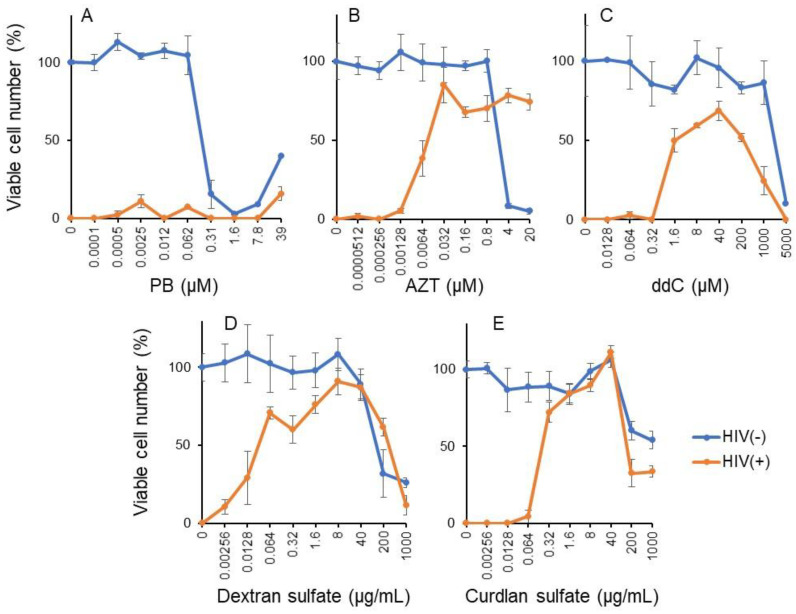
Background: Pyoktanin blue (PB) is used for staining tissues and cells, and it is applied in photodynamic therapy due to its potent bactericidal activity. However, clinical application of PB as an antiviral and antitumor agent has been limited due to its potent toxicity. For clinical application, the antitumor and antiviral activity as well as the neurotoxicity of PB were re-evaluated with a chemotherapeutic index. Methods: Tumor-specificity (TS) was determined by the ratio of CC50 against normal oral cells/oral squamous cell carcinoma (OSCC); neurotoxicity by that of normal oral/neuronal cells; antiviral activity by that of mock-infected/virus-infected cells; and potency-selectivity expression (PSE) by dividing TS by CC50 (OSCC). Results: Antitumor activity of PB (assessed by TS and PSE) was comparable with that of DXR and much higher than that of 5-FU and melphalan. PB induced caspase-3 activation and subG1 cell accumulation in an OSCC cell line (Ca9-22). PB and anticancer drugs showed comparable cytotoxicity against both neuronal cells and OSCC cell lines. PB showed no detectable anti-HIV/HSV activity, in contrast to reverse transferase inhibitors, sulfated glucans, and alkaline extract of leaves of S.P. Conclusions: PB showed first-class anticancer activity and neurotoxicity, suggesting the importance of establishing the safe treatment schedule.

Keywords: anti-HIV; anti-HSV; anticancer activity; apoptosis; caspase-3; chemotherapeutic index; oral cancer; pyoktanin; subG1 accumulation.


Conflict of interest statement

The authors declare no conflict of interest.

Figures

Figure 1
Figure 1
Structure of pyoktanin blue (PB) and design of the present study.
Figure 2
Figure 2
Dose–response curve of cytotoxicity of pyoktanin (PB) and three anticancer drugs (DXR, 5-FU and melphalan). Cells were incubated for 48 h with the indicated concentrations of test compounds. Experiments were repeated three times. Each value in each panel represents mean ± S.D. of triplicate assays. PB, pyoktanin blue; DXR, doxorubicin; 5-FU, fluorouracil.
Figure 3
Figure 3
Pyoktanin (PB) induces apoptosis in Ca9-22 cells. Ca9-22 cells were incubated for 24 h and then subjected to morphological observation under the light microscopy (A), Western blot analysis (B), and cell sorter analysis (C). The percentage of subG1 population was determined in triplicate and expressed as mean ± S.D. AD, actinomycin D; * statistically significant difference from control (p < 0.05).
Figure 4
Figure 4
Cytotoxicity of PB and anticancer drugs against PC12, SH-SY5Y, and LY-PPB6 (A) and differentiated PC12 cells (B). Each value represents mean ± S.D. of triplicate assays.
Figure 5
Figure 5
PB failed to induce anti-HIV activity. Mock and HIV-infected MT-4 were treated with the indicated concentrations of pyoktanin (A), AZT (B), ddC (C), dextran sulfate (D), and curdlan sulfate (E) for 5 days to determine the viability by MTT method. Each value represents mean ± S.D. of triplicate assay.
Figure 6
Figure 6
PB failed to induce anti-HSV activity. 100xHSV (MOI = 1) and the indicated concentrations of PB (A) or SE (B) were mixed for 3 min, diluted to x100 with culture medium to bring MOI = 0.01, added to Vero cells, and then incubated for 3 days (HSV(+)). Mock-infected Vero cells were treated for 3 min with the indicated concentrations of sample without virus, washed, and cultured for 3 days in fresh culture medium (HSV(−)) according to method 3 [24]. Viable cell number was then determined by MTT method. Each value represents mean ± S.D. of triplicate assay.

References

    1. Ghanadan A., Kamyab-Hesari K., Daneshpajouh M., Balighi K., Normohammadpour P. Nodular colloid degeneration of the skin: Report of three cases with review and update. Indian Dermatol. Online J. 2014;5:S36–S39. doi: 10.4103/2229-5178.144527. - DOI - PMC - PubMed
    1. Hayashi N., Sasaki T., Tomura N., Okada H., Kuwata T. Removal of a malignant cystic brain tumor utilizing pyoktanin blue and fibrin glue: Technical note. Surg. Neurol. Int. 2017;8:24. doi: 10.4103/2152-7806.200578. - DOI - PMC - PubMed
    1. Otani N., Wada K., Toyooka T., Takeuchi S., Tomiyama A., Mori K. Usefulness of dural surface tracing of the cortical vessels with indocyanine green videoangiography just prior to dural opening for various cerebrovascular diseases. Surg. Neurol. Int. 2017;8:201. doi: 10.4103/sni.sni_202_17. - DOI - PMC - PubMed
    1. Takamiya S., Seki T., Yamazaki K., Sasamori T., Houkin K. Intraoperative Visualization of a Spinal Arachnoid Cyst Using Pyoktanin Blue. World Neurosurg. 2018;109:18–23. doi: 10.1016/j.wneu.2017.09.031. - DOI - PubMed
    1. Tripathi N., Sapra A. StatPearls, StatPearls Publishing Copyright © 2021. StatPearls Publishing LLC.; Treasure Island, FL, USA: 2021. Gram Staining.