

Expression Profile of New Marker Genes Involved in Differentiation of Canine Adipose-Derived Stem Cells into Osteoblasts
- PMID: 34206369
- PMCID: PMC8269079
- DOI: 10.3390/ijms22136663
Expression Profile of New Marker Genes Involved in Differentiation of Canine Adipose-Derived Stem Cells into Osteoblasts
Abstract
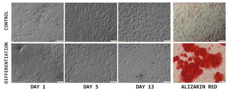
Next-generation sequencing (RNAseq) analysis of gene expression changes during the long-term in vitro culture and osteogenic differentiation of ASCs remains to be important, as the analysis provides important clues toward employing stem cells as a therapeutic intervention. In this study, the cells were isolated from adipose tissue obtained during routine surgical procedures and subjected to 14-day in vitro culture and differentiation. The mRNA transcript levels were evaluated using the Illumina platform, resulting in the detection of 19,856 gene transcripts. The most differentially expressed genes (fold change >|2|, adjusted p value < 0.05), between day 1, day 14 and differentiated cell cultures were extracted and subjected to bioinformatical analysis based on the R programming language. The results of this study provide molecular insight into the processes that occur during long-term in vitro culture and osteogenic differentiation of ASCs, allowing the re-evaluation of the roles of some genes in MSC progression towards a range of lineages. The results improve the knowledge of the molecular mechanisms associated with long-term in vitro culture and differentiation of ASCs, as well as providing a point of reference for potential in vivo and clinical studies regarding these cells' application in regenerative medicine.
Keywords: RNAseq; adipose; analysis; stem cells; transcriptome.
Conflict of interest statement
The authors declare no conflict of interest.
Figures


Similar articles
-
Identification and Characterization of Long Non-Coding RNAs in Osteogenic Differentiation of Human Adipose-Derived Stem Cells.Cell Physiol Biochem. 2017;42(3):1037-1050. doi: 10.1159/000478751. Epub 2017 Jun 28. Cell Physiol Biochem. 2017. PMID: 28662497
-
LncRNA-AK137033 inhibits the osteogenic potential of adipose-derived stem cells in diabetic osteoporosis by regulating Wnt signaling pathway via DNA methylation.Cell Prolif. 2022 Jan;55(1):e13174. doi: 10.1111/cpr.13174. Epub 2021 Dec 24. Cell Prolif. 2022. PMID: 34953002 Free PMC article.
-
Role of gender and anatomical region on induction of osteogenic differentiation of human adipose-derived stem cells.Ann Plast Surg. 2008 Mar;60(3):306-22. doi: 10.1097/SAP.0b013e3180621ff0. Ann Plast Surg. 2008. PMID: 18443514
-
Leporine-derived adipose precursor cells exhibit in vitro osteogenic potential.J Craniofac Surg. 2008 Mar;19(2):360-8. doi: 10.1097/SCS.0b013e318163e17b. J Craniofac Surg. 2008. PMID: 18362712
-
Effect of anatomical origin and cell passage number on the stemness and osteogenic differentiation potential of canine adipose-derived stem cells.Stem Cell Rev Rep. 2012 Dec;8(4):1211-22. doi: 10.1007/s12015-012-9397-0. Stem Cell Rev Rep. 2012. PMID: 22773405
Cited by
-
Differential regulation of apoptosis-related genes during long-term culture and differentiation of canine adipose-derived stem cells - a functional bioinformatical analysis.Front Genet. 2025 Jan 6;15:1515778. doi: 10.3389/fgene.2024.1515778. eCollection 2024. Front Genet. 2025. PMID: 39834550 Free PMC article.
-
Polypeptide N-acetylgalactosaminyltransferase-15 regulates adipogenesis in human SGBS cells.Sci Rep. 2024 Aug 29;14(1):20049. doi: 10.1038/s41598-024-70930-5. Sci Rep. 2024. PMID: 39209927 Free PMC article.
-
Expression Profile of New Gene Markers Involved in Differentiation of Canine Adipose-Derived Stem Cells into Chondrocytes.Genes (Basel). 2022 Sep 16;13(9):1664. doi: 10.3390/genes13091664. Genes (Basel). 2022. PMID: 36140831 Free PMC article.
-
Bone Metastasis Challenge: New Ideas and Future.Int J Mol Sci. 2023 Mar 24;24(7):6161. doi: 10.3390/ijms24076161. Int J Mol Sci. 2023. PMID: 37047132 Free PMC article.
References
-
- Moncrieff L., Mozdziak P., Jeseta M., Machatkova M., Kranc W., Kempisty B. Ovarian follicular cells—Living in the shadow of stemness cellular competence. Med. J. Cell Biol. 2019;7:134–140. doi: 10.2478/acb-2019-0018. - DOI
-
- Kulus M., Kulus J., Jankowski M., Borowiec B., Jeseta M., Bukowska D., Brüssow K.P., Kempisty B., Antosik P. The use of mesenchymal stem cells in veterinary medicine. Med. J. Cell Biol. 2018;6:101–107. doi: 10.2478/acb-2018-0016. - DOI
-
- Stefańska K., Sibiak R., Hutchings G., Dompe C., Moncrieff L., Janowicz K., Jeseta M., Kempisty B., Machatkova M., Mozdziak P. Evidence for existence of molecular stemness markers in porcine ovarian follicular granulosa cells. Med. J. Cell Biol. 2019 doi: 10.2478/acb-2019-0025. - DOI
MeSH terms
Grants and funding
LinkOut - more resources
Full Text Sources
Medical
