. 2021 Jun 26;26(13):3915.
doi: 10.3390/molecules26133915.

Novel Bradykinin Receptor Inhibitors Inhibit Proliferation and Promote the Apoptosis of Hepatocellular Carcinoma Cells by Inhibiting the ERK Pathway

Affiliations

Novel Bradykinin Receptor Inhibitors Inhibit Proliferation and Promote the Apoptosis of Hepatocellular Carcinoma Cells by Inhibiting the ERK Pathway

Yiou Wang et al. Molecules. .

Abstract

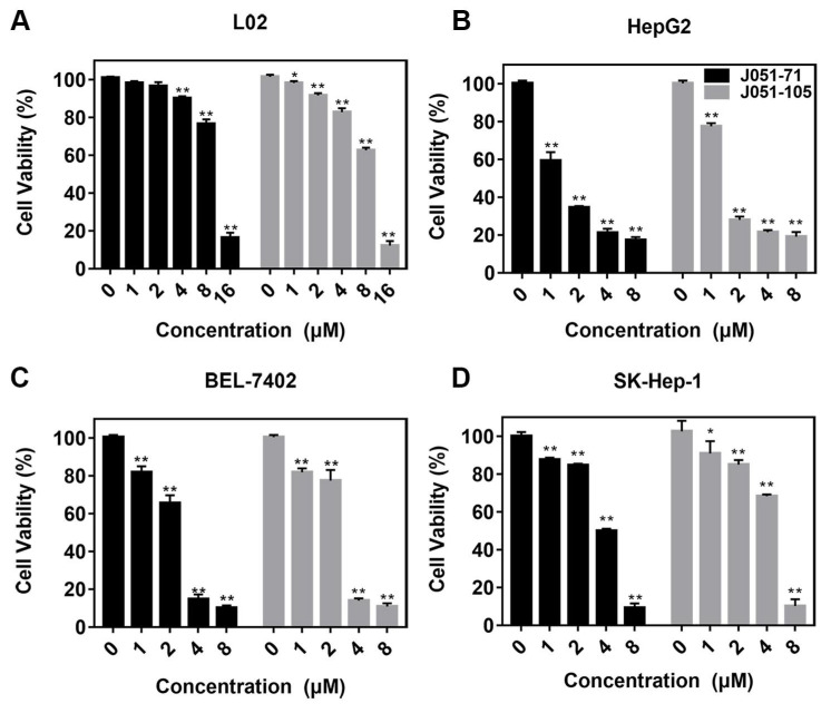
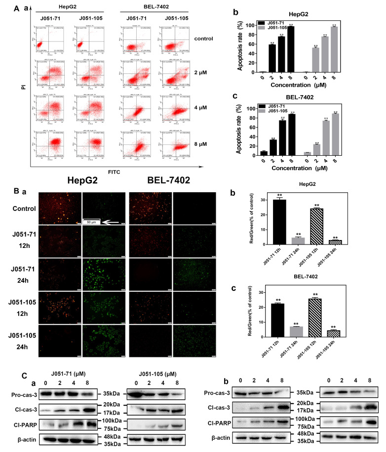
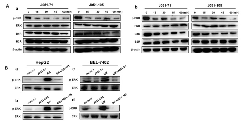
Hepatocellular carcinoma (HCC) is the fifth most common cancer worldwide. Studies have shown that bradykinin (BK) is highly expressed in liver cancer. We designed the novel BK receptor inhibitors J051-71 and J051-105, which reduced the viability of liver cancer cells and inhibited the formation of cancer cell colonies. J051-71 and J051-105 reduced cell proliferation and induced apoptosis in HepG2 and BEL-7402 cells, which may be due to the inhibition of the extracellular regulated protein kinase (ERK) signaling pathway. In addition, these BK receptor inhibitors reversed the cell proliferation induced by BK in HepG2 and BEL-7402 cells by downregulating B1 receptor expression. Inhibiting B1 receptor expression decreased the protein levels of p-ERK and reduced the malignant progression of HCC, providing a potential target for HCC therapy.

Keywords: ERK signaling pathway; apoptosis; bradykinin B1 receptor; bradykinin receptor inhibitor; hepatocellular carcinoma.


Conflict of interest statement

The authors declare no conflict of interest with the contents of this article.

Figures

Figure 1
Figure 1
Synthesis of bradykinin receptor inhibitors. (A) Structural formula of J051-71. (B) NMR hydrogen spectrum of J051-71. (C) Structural formula of J051-105. (D) NMR hydrogen spectrum of J051-105.
Figure 2
Figure 2
Bradykinin receptor inhibitors inhibit the cell viability of primary hepatocellular carcinoma cells. (A) L02, (B) HepG2, (C) BEL-7402, and (D) SK-Hep-1 cells were treated with various concentrations (0, 1, 2, 4, and 8 μM) of J051-71 or J051-105 for 24 h. The cell viability of hepatocellular carcinoma cells was determined by the MTT assay (n = 3). * p < 0.05, ** p < 0.01: the test concentrations (1, 2, 4, and 8 μM) vs. the test concentration (0 μM).
Figure 3
Figure 3
Bradykinin receptor inhibitors inhibit the proliferation of hepatocellular carcinoma cells. (A) (a) Colonies were stained with crystal violet to evaluate the cell colony formation of HepG2 and BEL-7402 cells. (b) Colony formation assay of HepG2 cells treated with various concentrations (0.5 and 1 μM) J051-71 and J051-105 (n = 3), HepG2 cells without treatment were used as control. * p < 0.05, ** p < 0.01 the test concentrations (0.5 and 1 μM) vs. control, respectively. (B) (a) Colonies were stained with crystal violet to evaluate the cell colony formation of BEL-7402 cells. (b) Colony formation assay of BEL-7402 cells treated with various concentrations (0.5 and 1 μM) J051-71 and J051-105 (n = 3), BEL-7402 cells without treatment were used as control. * p < 0.05, ** p < 0.01 the test concentrations (0.5 and 1 μM) vs. control, respectively. (C) (a) Cells were treated with BK at a concentration of 1 μM and J051-71 and J051-105 at 1 μM. (b) Colony formation assay of HepG2 and BEL-7402 cells treated with BK, BK and J051-71 or BK and J051-105 (n = 3), HepG2 and BEL-7402 cells without BK treatment were used as control. * p < 0.05, ** p < 0.01: BK and J051-71 or BK and J051-105 vs. BK. # p < 0.05: BK vs. control.
Figure 4
Figure 4
Bradykinin receptor inhibitors promote the apoptosis of hepatocellular carcinoma. (A) HepG2 and BEL-7402 cells were exposed to J051-71 or J051-105 (0, 2, 4, and 8 μM) for 24 h (n = 3). (a). BK inhibitors affect the outcome of hepatocellular carcinoma cell apoptosis. Quantification of apoptotic HepG2 (b) and BEL-7402 (c) cells, ** p < 0.01: the test concentrations (2, 4, and 8 μM) vs. the test concentration (0 μM). (B) HepG2 and BEL-7402 cells were exposed to 2 μM J051-71 alone or 2 μM J051-105 alone for 12 h or 24 h (n = 3). (a). Qualitative results of fluorescence microscopy of mitochondrial membrane potential. Apoptotic cells were identified by JC-1 staining (green), and non-apoptotic cells were identified by JC-1 staining (red). Scale bars are 50 μm. The calculated percentage ratio of red to green for HepG2 (b) and BEL-7402 (c) cells treated with J051-71 and J051-105 for 12h and 24h (n = 10), ** p < 0.01 vs. control, respectively. (C) Western blot analysis of the protein levels of pro-caspase 3, cleaved caspase-3, and cleaved PARP in HepG2 (a) and BEL-7402 (b) cells (n = 3). β-actin served as the loading control.
Figure 5
Figure 5
Bradykinin receptor inhibitors reduce the proliferation of hepatocellular carcinoma cells by inhibiting the activation of the ERK signaling pathway. (A) HepG2 (a) and BEL-7402 (b) cells were treated with 2 μM J051-71 and J051-105 for 0, 15, 30, 45, and 60 min (n = 3). Protein levels of B1R, B2R, p-ERK, and ERK were determined by Western blot. β-actin served as the loading control. (B) Western blot expression of the indicated proteins in HepG2 cells treated with 2 μM J051-71 (a) and J051-105 (b) or BEL-7402 cells treated with J051-71 (c) and J051-105 (d) for 30 min (n = 3).

Similar articles

Cited by

References

    1. Shi J., Lai E.C., Li N., Guo W.X., Xue J., Lau W.Y., Wu M.C., Cheng S.Q. Surgical treatment of hepatocellular carcinoma with portal vein tumor thrombus. Ann. Surg. Oncol. 2010;7:2073–2080. doi: 10.1245/s10434-010-0940-4. - DOI - PubMed
    1. Yang Y., Nagano H., Ota H., Morimoto O., Nakamura M., Wada H., Node T., Damdinsuren B., Marubashi S., Miyamoto A., et al. Patterns and clinicopathologic features of extrahepatic recurrence of hepatocellular carcinoma after curative resection. Surgery. 2007;141:196–202. doi: 10.1016/j.surg.2006.06.033. - DOI - PubMed
    1. Singh A.K., Kumar R., Pandey A.K. Hepatocellular Carcinoma: Causes, Mechanism of Progression and Biomarkers. Curr. Chem. Genom. Transl. Med. 2018;12:9–26. doi: 10.2174/2213988501812010009. - DOI - PMC - PubMed
    1. Hartke J., Johnson M., Ghabril M. The diagnosis and treatment of hepatocellular carcinoma. Semin. Diagn. Pathol. 2017;34:153–159. doi: 10.1053/j.semdp.2016.12.011. - DOI - PubMed
    1. Kudo M., Finn R.S., Qin S., Han K.H., Ikeda K., Piscaglia F., Baron A., Park J.-W., Han G., Jassem J., et al. Lenvatinib versus sorafenib in first-line treatment of patients with unresectable hepatocellular carcinoma: A randomised phase 3 non-inferiority trial. Lancet. 2018;391:1163–1173. doi: 10.1016/S0140-6736(18)30207-1. - DOI - PubMed

MeSH terms

Substances

LinkOut - more resources