. 2021 Jun 26;26(13):3923.
doi: 10.3390/molecules26133923.

Discovery of New Pyrazolopyridine, Furopyridine, and Pyridine Derivatives as CDK2 Inhibitors: Design, Synthesis, Docking Studies, and Anti-Proliferative Activity

Affiliations

Discovery of New Pyrazolopyridine, Furopyridine, and Pyridine Derivatives as CDK2 Inhibitors: Design, Synthesis, Docking Studies, and Anti-Proliferative Activity

Adel A-H Abdel-Rahman et al. Molecules. .

Abstract

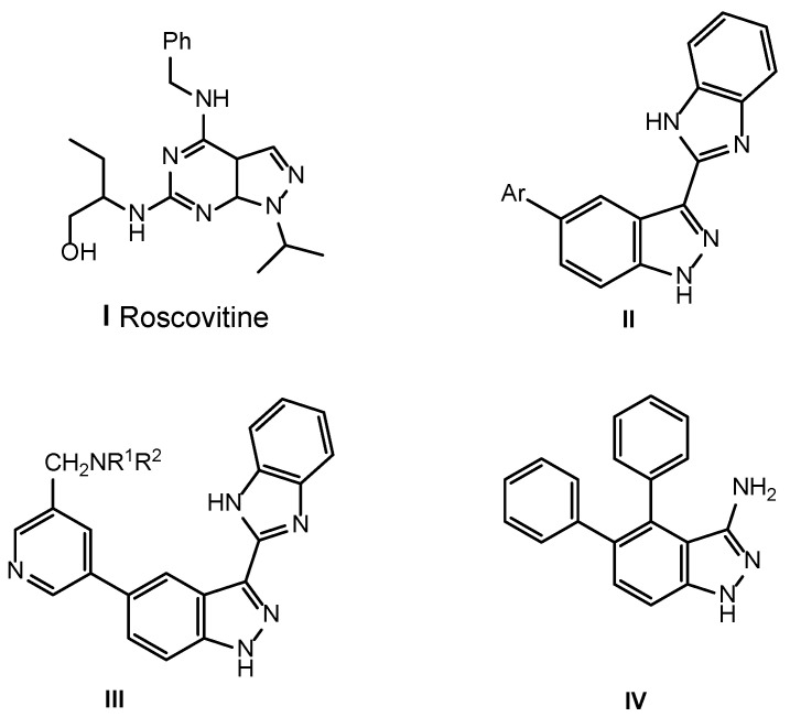
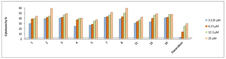
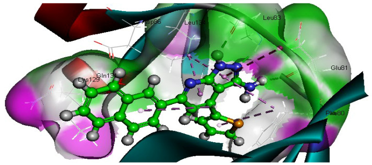
New pyridine, pyrazoloyridine, and furopyridine derivatives substituted with naphthyl and thienyl moieties were designed and synthesized starting from 6-(naphthalen-2-yl)-2-oxo-4-(thiophen-2-yl)-1,2-dihydropyridine-3-carbonitrile (1). The chloro, methoxy, cholroacetoxy, imidazolyl, azide, and arylamino derivatives were prepared to obtain the pyridine--C2 functionalized derivatives. The derived pyrazolpyridine-N-glycosides were synthesized via heterocyclization of the C2-thioxopyridine derivative followed by glycosylation using glucose and galactose. The furopyridine derivative 14 and the tricyclic pyrido[3',2':4,5]furo[3,2-d]pyrimidine 15 were prepared via heterocyclization of the ester derivative followed by a reaction with formamide. The newly synthesized compounds were evaluated for their ability to in vitro inhibit the CDK2 enzyme. In addition, the cytotoxicity of the compounds was tested against four different human cancer cell lines (HCT-116, MCF-7, HepG2, and A549). The CDK2/cyclin A2 enzyme inhibitory results revealed that pyridone 1, 2-chloro-6-(naphthalen-2-yl)-4-(thiophen-2-yl)nicotinonitrile (4), 6-(naphthalen-2-yl)-4-(thiophen-2-yl)-1H-pyrazolo[3,4-b]pyridin-3-amine (8), S-(3-cyano-6-(naphthaen-2-yl)-4-(thiophen-2-yl)pyridin-2-yl) 2-chloroethanethioate (11), and ethyl 3-amino-6-(naphthalen-2-yl)-4-(thiophen-2-yl)furo[2,3-b]pyridine-2-carboxylate (14) are among the most active inhibitors with IC50 values of 0.57, 0.24, 0.65, 0.50, and 0.93 µM, respectively, compared to roscovitine (IC50 0.394 μM). Most compounds showed significant inhibition on different human cancer cell lines (HCT-116, MCF-7, HepG2, and A549) with IC50 ranges of 31.3-49.0, 19.3-55.5, 22.7-44.8, and 36.8-70.7 μM, respectively compared to doxorubicin (IC50 40.0, 64.8, 24.7 and 58.1 µM, respectively). Furthermore, a molecular docking study suggests that most of the target compounds have a similar binding mode as a reference compound in the active site of the CDK2 enzyme. The structural requirements controlling the CDK2 inhibitory activity were determined through the generation of a statistically significant 2D-QSAR model.

Keywords: CDK2; HepG2; anticancer; docking; imidazole; pyrazolo[3,4-b]pyridine; pyridine.


Conflict of interest statement

The authors declare that they have no conflict of interest.

Figures

Figure 1
Figure 1
Roscovitine, (pyridin-3-yl)-1H-indazole and imidazolyl-1H-indazole derivatives with potent inhibition activity for CDK2.
Scheme 1
Scheme 1
Synthesis of substituted pyridine derivatives (2, 4-6) and (imidazolyl)oxynicotinonitrile derivatives 3.
Scheme 2
Scheme 2
Synthesis of pyrazolopyridine-N-glycosides (9 and 10) and pyridinyl-2- azidoethanethioate derivative 12.
Scheme 3
Scheme 3
Synthesis of substituted pyrido[3′,2′:4,5]furo[3,2-d]pyrimidin-4(3H)-one derivative 15.
Figure 2
Figure 2
Dose-dependent cytotoxic activities of the compounds against HCT-116 cancer cells according to the MTT assay.
Figure 3
Figure 3
Dose-dependent cytotoxic activities of the compounds against MCF-7 cancer cells according to the MTT assay.
Figure 4
Figure 4
Dose-dependent cytotoxic activities of the compounds against HepG2 cancer cells according to the MTT assay.
Figure 5
Figure 5
Dose-dependent cytotoxic activities of the compounds against A549 cancer cells according to the MTT assay.
Figure 6
Figure 6
Two-dimensional (2D) diagram of roscovitine in the active site of CDK2. Dot green line represents the hydrogen bond donor and acceptor. Orange color line represents the hydrophobic interaction.Amino acids with green color represent the Van der Waals interaction, while the purple amino acids represent the electrostatic interaction.
Figure 7
Figure 7
Two-dimensional (2D) and 3D docking of compound 1 into the CDK2 active site. Amino acids with green color represent the Van der Waals interaction; the purple amino acids represent the electrostatic interaction, while the blue amino acid represents the hydrophobic interaction.
Figure 7
Figure 7
Two-dimensional (2D) and 3D docking of compound 1 into the CDK2 active site. Amino acids with green color represent the Van der Waals interaction; the purple amino acids represent the electrostatic interaction, while the blue amino acid represents the hydrophobic interaction.
Figure 8
Figure 8
Docking of compound 4 into the CDK2 active site. Purple color represent carbon atoms, gray color represent hydrogen atom, blue color represent nitrogen atom, orange color represent sulfur atom and green color represent chlorine atom.
Figure 9
Figure 9
Docking of compound 8 into the CDK2 active site. Gray color represent carbon atoms, blue color represent nitrogen atoms, while the orange color represent sulfur atoms.
Figure 9
Figure 9
Docking of compound 8 into the CDK2 active site. Gray color represent carbon atoms, blue color represent nitrogen atoms, while the orange color represent sulfur atoms.
Figure 10
Figure 10
Docking of compound 11 into the CDK2 active site. Red color represent the carbon atoms of molecules, gray color represent hydrogen atoms, the blue color represent the nitrogen atoms, the orange color represent sulfur atom and The green color represent chlorine atom.
Figure 11
Figure 11
Docking of compound 14 into the CDK2 active site.
Figure 12
Figure 12
Alignment of compound 11 (green) with roscovitine (purple).
Figure 13
Figure 13
Correlation between the binding energy and the inhibitory profile of the investigated molecules.
Figure 14
Figure 14
MLR-QSAR model plot of correlations representing the observed versus predicted IC50 μM values for the compounds 15, 7, 11, 13, and 14 against the CDK2 enzyme.
Figure 15
Figure 15
External test set used in validation of generated QSAR model.

References

    1. Alghassimou D., Claude P. The serine/threonine kinases that control cell cycle progression as therapeutic targets. Bull. Cancer. 2011;98:1335–1345. - PubMed
    1. Johnsona L.N., De Moliner E., Browna N.R., Songa H., Barfordc D., Endicotta J.A., Noble M.E. structure studies with inhibitors of the cell cycle regulatory kinase cyclin-dependent protein kinase 2. Pharmacol. Ther. 2002;93:113–124. doi: 10.1016/S0163-7258(02)00181-X. - DOI - PubMed
    1. Chen J., Pang L., Wang W., Wang L., Zhang J.Z., Zhu T. Decoding molecular mechanism of inhibitor bindings to CDK2 using molecular dynamics simulations and binding free energy calculations. J. Biomol. Struct. Dyn. 2020;38:985–996. doi: 10.1080/07391102.2019.1591304. - DOI - PubMed
    1. Chen J., Wang X., Zhang J.Z., Zhu T. effect of substitution in different positions of aminothiazole hing-binding scaffolds on inhibitor-CDK2 association probed by interaction entropy method. ACS Omega. 2018;3:18052–18064. doi: 10.1021/acsomega.8b02354. - DOI
    1. Kontopidis G., McInnes C., Pandalaneni S.R., McNae I., Gibson D., Mezna M., Thomas M., Wood G., Wang S., Walkinshaw M.D., et al. Differential binding of inhibitors to active and inactive CDK2 provides insights for drug design. Chem. Biol. 2006;13:201–211. doi: 10.1016/j.chembiol.2005.11.011. - DOI - PubMed

MeSH terms

LinkOut - more resources