. 2021 Jun 26;13(7):1246.
doi: 10.3390/v13071246.

Human Defensins Inhibit SARS-CoV-2 Infection by Blocking Viral Entry

Affiliations

Human Defensins Inhibit SARS-CoV-2 Infection by Blocking Viral Entry

Chuan Xu et al. Viruses. .

Abstract

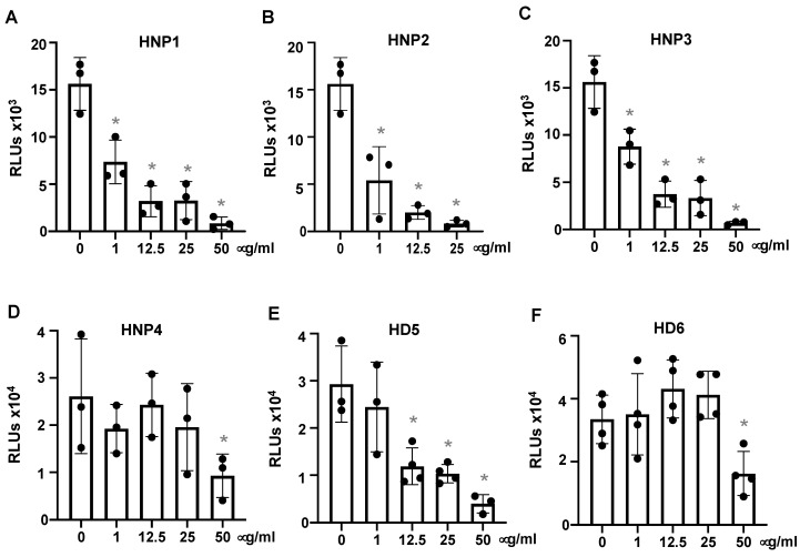
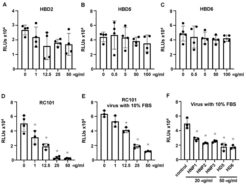
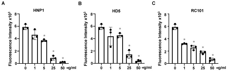
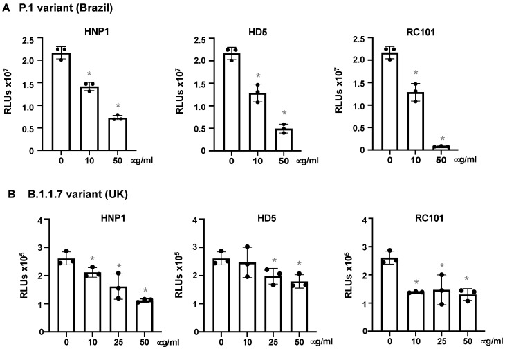
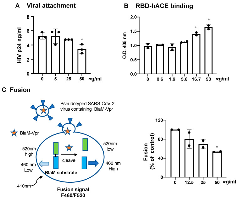
Innate immunity during acute infection plays a critical role in the disease severity of severe acute respiratory syndrome (SARS) and Middle East respiratory syndrome (MERS), and is likely to contribute to COVID-19 disease outcomes. Defensins are highly abundant innate immune factors in neutrophils and epithelial cells, including intestinal Paneth cells, and exhibit antimicrobial and immune-modulatory activities. In this study, we investigated the effects of human α- and β-defensins and RC101, a θ-defensin analog, on SARS-CoV-2 infection. We found that human neutrophil peptides (HNPs) 1-3, human defensin (HD) 5 and RC101 exhibited potent antiviral activity against pseudotyped viruses expressing SARS-CoV-2 spike proteins. HNP4 and HD6 had weak anti-SARS-CoV-2 activity, whereas human β-defensins (HBD2, HBD5 and HBD6) had no effect. HNP1, HD5 and RC101 also inhibited infection by replication-competent SARS-CoV-2 viruses and SARS-CoV-2 variants. Pretreatment of cells with HNP1, HD5 or RC101 provided some protection against viral infection. These defensins did not have an effect when provided post-infection, indicating their effect was directed towards viral entry. Indeed, HNP1 inhibited viral fusion but not the binding of the spike receptor-binding domain to hACE2. The anti-SARS-CoV-2 effect of defensins was influenced by the structure of the peptides, as linear unstructured forms of HNP1 and HD5 lost their antiviral function. Pro-HD5, the precursor of HD5, did not block infection by SARS-CoV-2. High virus titers overcame the effect of low levels of HNP1, indicating that defensins act on the virion. HNP1, HD5 and RC101 also blocked viral infection of intestinal and lung epithelial cells. The protective effects of defensins reported here suggest that they may be useful additives to the antivirus arsenal and should be thoroughly studied.

Keywords: SARS-CoV-2; antimicrobial peptides; defensins.


Conflict of interest statement

The authors declare that there is no conflict of interest.

Figures

Figure 1
Figure 1
Human α-defensins inhibit infection by pseudotyped SARS-CoV-2 virus. Pseudotyped luciferase reporter virus expressing SARS-CoV-2 S protein was incubated with or without HNPs1-4, HD5 or HD6 at 37 °C for 1 h. Viruses with or without defensin treatment were used to infect HEK293T cells expressing hACE2 (AF) as described in the Methods. Infected cells were cultured for three days before measuring luciferase activity. The significance of the differences between defensin-treated virions and mocked-treated controls was calculated by Student’s two-tailed, unpaired t = test; * p < 0.05. Data are means ± SD and are representative of four independent experiments.
Figure 2
Figure 2
Theta defensin analog RC101 but not HBDs inhibits pseudotyped SARS-CoV-2 infection. Pseudotyped luciferase reporter viruses expressing SARS-CoV-2 S proteins were incubated with or without the indicated concentrations of HBD2, HBD5, HBD6 or RC101 at 37 °C for 1 h followed by infection of HEK293T cells expressing hACE2 (AD) as described in the Methods. Infected cells were cultured for three days before measuring luciferase activity. The effect of defensins on the virus in the presence of 10% FBS was also determined (E,F). Differences between defensin-treated virions and non-treated controls were calculated by Student’s two-tailed, unpaired t-test; * p < 0.05. Data are means ± SD and are representative of three independent experiments.
Figure 3
Figure 3
Defensins inhibit infection by replication-competent SARS-CoV-2 viruses. Replication-competent SARS-CoV-2 viruses expressing mNeonGreen (at MOI of five) were incubated with HNP1 (A), HD5 (B) and RC101 (C) at different concentrations and at 37 °C for 1 h before addition to Vero E6 cells for 2 h. Infected cells were then cultured in FluoroBrite media with 10% FBS for two days. The fluorescence from productive viral infections was measured using Biotek Cytation 5. The differences between defensin-treated virions and non-treated controls were calculated by Student’s two-tailed, unpaired t-test; * p < 0.05. Data are means ± SD and are representative of three independent experiments.
Figure 4
Figure 4
HNP1, HD5 and RC101 inhibit infection by pseudotyped SARS-CoV-2 variants. Pseudotyped luciferase reporter virus expressing SARS-CoV-2 S protein from the P.1 variant (A) or the B.1.1.7 variant (B) was incubated with or without HNP1, HD5 and RC101 at 37 °C for 1 h. Viruses were added to HeLa-hACE2 cells and incubated at 37 °C for 2 h. Infected cells were cultured for two days before measuring luciferase activity. The significance of the differences between defensin-treated virions and mocked-treated controls was calculated by Student’s two-tailed, unpaired t-test; * p < 0.05. Data are means ± SD and are representative of three independent experiments.
Figure 5
Figure 5
Defensins are ineffective after viral entry. (A) To determine the effect of defensins on target cell resistance to infection, HEK293T-hACE2 cells were incubated with defensins in the presence of FBS for 1 h, and then exposed to pseudotyped SARS-CoV-2 luciferase reporter virus without defensins for 2 h. Cells were washed and then cultured in fresh media for three days before measuring luciferase activity. (B) To determine the post-entry effect of defensins, HEK293T-hACE2 cells were infected with pseudotyped SARS-CoV-2 viruses for 2 h followed by treatment of infected cells with the indicated concentration of defensins. The differences between defensin-treated samples and untreated controls were calculated by Student’s two-tailed t-test; * p < 0.05. Data are means ± SD of triplicate samples and are representative of three independent experiments.
Figure 6
Figure 6
The effect of HNP1 on viral attachment, spike RBD-hACE2 interaction and viral fusion. (A) Pseudotyped SARS-CoV-2 viruses were treated with HNP1 at different concentrations for 1 h at 37 °C. The virus-defensin mixture was added to HeLa-hACE2 cells, and cells were incubated at 4 °C for 2 h. After washing off unbound viruses, cells were lysed, and cell-associated HIVp24 was determined as described in the Methods. (B) Immobilized spike RBD proteins on the plate were incubated with biotinylated hACE proteins that were pre-treated with defensins at different concentrations for 1 h. After incubation at 37 °C for 1 h, the plate was washed and the bound hACE2 proteins were detected by incubation with alkylate phosphatase (AP)-conjugated streptavidin followed by the AP colorimetric assay as described in the Methods. For panels A and B, the differences between defensin-treated samples and untreated controls were calculated by Student’s two-tailed t-test; * p < 0.05. Data are means ± SD of triplicate samples and are representative of three independent experiments. (C) A schematic of Förster resonance energy transfer (FRET)-based BlaM assay is shown on the left panel. Pseudotyped SARS-CoV-2 viruses containing BlaM-Vpr proteins were incubated with Huh7.5-ACE2-TMPRSS2 cells at 16 °C for 30 min at 1550× g. After washing off unbound viruses, cells were treated with HNP1 at different concentrations and incubated at 37 °C for 2 h for viral fusion. The BlaM substrate, CCF4-AM, was added to cells, which were incubated at 11oC overnight. The cytoplasmic BlaM activity from viral fusion was determined by measuring the ratio of blue (460 nm) to green (520 nm) fluorescence with excitation at 410 nm. The differences between defensin-treated samples and untreated controls were calculated by Student’s two-tailed t-test; * p < 0.05. Data are means ± SD of two independent experiments.
Figure 7
Figure 7
The structure of defensins required for anti-SARS-CoV-2 activity. To determine the effect of linear defensins (A) or proHD5 (B) on viral infection, pseudotyped SARS-CoV-2 virus was incubated with indicated concentrations of [Abu]HNP1 or [Abu]HD5 for 1 h before infection of HEK293T-hACE2 cells. Infected cells were cultured for three days before measuring luciferase activity. There was no difference between defensin-treated samples and untreated controls as calculated by Student’s two-tailed t-test. Data are means ± SD of triplicate samples and are representative of two independent experiments.
Figure 8
Figure 8
The effect of viral titers and cell types on defensin-mediated SARS-CoV-2 inhibition. (A) To determine the effect of virus titers on the antiviral activity of HNP1, indicated dilutions of serum-free pseudotyped SARS-CoV-2 viruses were incubated with 1 μg/mL (left) or 25 μg/mL (right) HNP1 at 37 °C for 1 h before infection of HEK293T-hACE2 cells. The percentage of control was calculated using the formula: (RLU of defensin-treated samples/RLUs of average of untreated samples at the same virus titer) × 100. (B) To determine the effect of defensins on SARS-CoV-2 infection of intestinal and lung epithelial cells expressing endogenous hACE2 receptors, pseudotyped SARS-CoV-2 viruses were incubated with 25 μg/mL HNP1, HD5 or RC101 at 37 °C for 1 h, then added to intestinal CaCo-2 cells (left) or lung epithelial A549 cells (right) at 37 °C for 1.5 h. After washing off unbound viruses, infected cells were cultured for three days before measuring luciferase activities. The differences between defensin-treated samples and untreated controls were calculated using Student’s two-tailed t-test; * p < 0.05. Data are means ± SD of triplicate samples and are representative of three independent experiments.

References

    1. [(accessed on 23 June 2021)]; Available online: https://coronavirus.jhu.edu/map.html.
    1. Park A., Iwasaki A. Type I and Type III Interferons—Induction, Signaling, Evasion, and Application to Combat COVID-19. Cell Host Microbe. 2020;27:870–878. doi: 10.1016/j.chom.2020.05.008. - DOI - PMC - PubMed
    1. Channappanavar R., Fehr A.R., Vijay R., Mack M., Zhao J., Meyerholz D.K., Perlman S. Dysregulated Type I Interferon and Inflammatory Monocyte-Macrophage Responses Cause Lethal Pneumonia in SARS-CoV-Infected Mice. Cell Host Microbe. 2016;19:181–193. doi: 10.1016/j.chom.2016.01.007. - DOI - PMC - PubMed
    1. Channappanavar R., Fehr A.R., Zheng J., Wohlford-Lenane C., Abrahante J.E., Mack M., Sompallae R., McCray P.B., Jr., Meyerholz D.K., Perlman S. IFN-I response timing relative to virus replication determines MERS coronavirus infection outcomes. J. Clin. Investig. 2019;129:3625–3639. doi: 10.1172/JCI126363. - DOI - PMC - PubMed
    1. Ganz T. Defensins: Antimicrobial peptides of innate immunity. Nat. Rev. Immunol. 2003;3:710–720. doi: 10.1038/nri1180. - DOI - PubMed

Publication types