. 2021 Jun 18;22(12):6537.
doi: 10.3390/ijms22126537.

Expression Profiling and Bioinformatics Analysis of CircRNA in Mice Brain Infected with Rabies Virus

Affiliations

Expression Profiling and Bioinformatics Analysis of CircRNA in Mice Brain Infected with Rabies Virus

Wen Zhao et al. Int J Mol Sci. .

Abstract

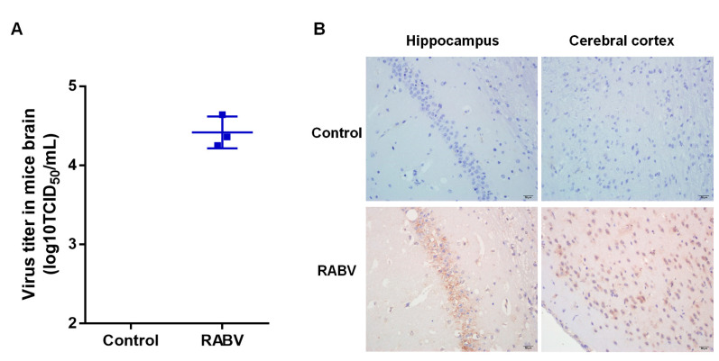
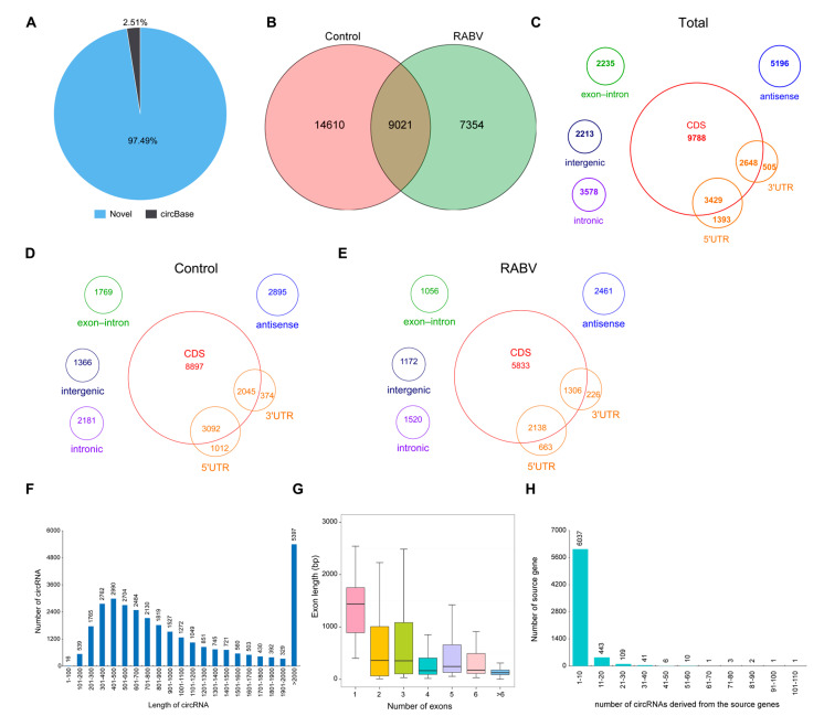
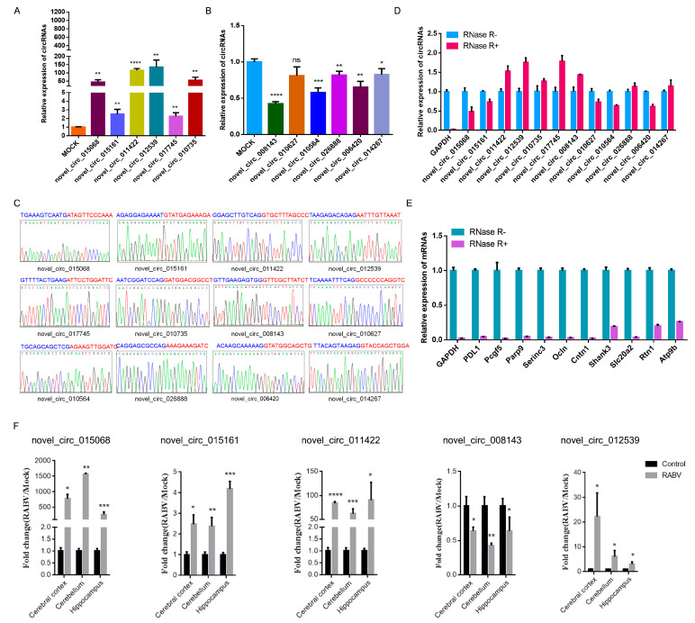
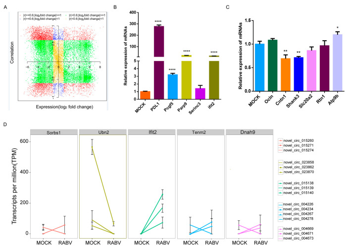
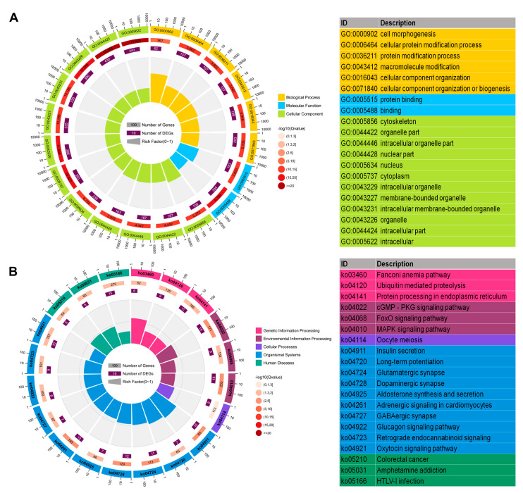
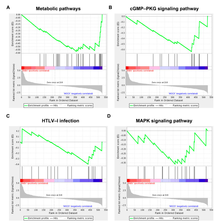
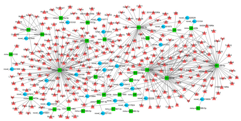
Rabies virus (RABV) induces acute, fatal encephalitis in mammals including humans. The circRNAs are important in virus infection process, but whether circRNAs regulated RABV infection remains largely unknown. Here, mice brain with or without the RABV CVS-11 strain were subjected to RNA sequencing and a total of 30,985 circRNAs were obtained. Among these, 9021 candidates were shared in both groups, and 14,610 and 7354 circRNAs were expressed specifically to the control and experimental groups, indicating that certain circRNAs were specifically inhibited or induced on RABV infection. The circRNAs mainly derived from coding exons. In total, 636 circRNAs were differentially expressed in RABV infection, of which 426 significantly upregulated and 210 significantly downregulated (p < 0.05 and fold change ≥2). The expression of randomly selected 6 upregulated and 6 downregulated circRNAs was tested by RT-qPCR, and the expression trend of the 11 out of 12 circRNAs was consistent in RT- qPCR and RNA-seq analysis. Rnase R-resistant assay and Sanger sequencing were conducted to verify the circularity of circRNAs. GO analysis demonstrated that source genes of all differentially regulated circRNAs were mainly related to cell plasticity and synapse function. Both KEGG and GSEA analysis revealed that these source genes were engaged in the cGMP-PKG and MAPK signaling pathway, and HTLV-I infection. Also, pathways related to glucose metabolism and synaptic functions were enriched in KEGG analysis. The circRNA-miRNA-mRNA network was built with 25 of 636 differentially expressed circRNAs, 264 mRNAs involved in RABV infection, and 29 miRNAs. Several miRNAs and many mRNAs in the network were reported to be related to viral infection and the immune response, suggesting that circRNAs could regulate RABV infection via interacting with miRNAs and mRNAs. Taken together, this study first characterized the transcriptomic pattern of circRNAs, and signaling pathways and function that circRNAs are involved in, which may indicate directions for further research to understand mechanisms of RABV pathogenesis.

Keywords: ceRNA network; circRNAs; enrichment analysis; mice brain; rabies virus.


Conflict of interest statement

The authors declare no conflict of interest.

Figures

Figure 1
Figure 1
Establishment of RABV mouse infection model. (A) Quantification of infectious virus titer in mice brain by indirect immunofluorescence assay (IFA); (B) Immunohistochemistry (IHC) analysis of the cerebral cortex and hippocampus to visualize RABV infection using anti-N antibody. Scale bar = 20 µm.
Figure 2
Figure 2
Profiling of circular RNAs in RABV- and DMEM-infected mice brains. (A) Pie chart showing the number of known circRNAs in Circbase and the new identified circRNAs; (B) The total number of circRNAs detected in the experimental and control groups; (C) Genomic features of circRNAs in both groups; (D) Genomic features of circRNAs in the control group; (E) Genomic features of circRNAs in the experimental group; (F) Illustration of the identified circRNAs length; (G) Box plot illustrating the length of exon that circularizes into circRNAs; (H) Bar plot showing that one gene could generate multiple circRNAs.
Figure 3
Figure 3
Validation of circRNAs. Expression changes of six upregulated (A) and six downregulated (B) circRNAs in RNA sequencing data were tested with divergent primers by RT-PCR; (C) Validation of back-splice junction sequences by Sanger sequencing; RNase-R resistance assay of 12 selected circRNAs (D) and corresponding linear transcripts (E); (F) Expression level of 5 circRNAs in the cerebral cortex, cerebellum, and hippocampus with or without RABV infection (p < 0.05, *; p < 0.01, **; p < 0.0005, ***; p < 0.0001, ****).
Figure 4
Figure 4
Expression correlation between circRNAs and linear counterparts. (A) The expression correlation between circRNAs and corresponding linear transcripts. The circRNAs are colored according to the Pearson correlation coefficient (r) and their expression pattern in RABV infection; (B) Expression changes of linear transcripts of six upregulated circRNAs and six downregulated circRNAs (C) were tested by RT-qPCR; (D) Variable expression patterns of circRNAs derived from Ubn2, Ifit2, Sorbs1, Tenm2, and Dnah9 (p < 0.05, *; p < 0.01, **; p < 0.0001, ****).
Figure 5
Figure 5
GO and KEGG enrichment analysis. The circular graph shows the top 20 enriched GO terms (A) and KEGG pathways (B) of source genes of 636 differentially expressed circRNAs.
Figure 6
Figure 6
Representative 4 significantly enriched gene sets from GSEA analysis. Source genes of significantly differentially expressed circRNAs were specifically engaged in (A) metabolic pathways, (B) the cGMP–PKG signaling pathway, (C) HTLV-I infection, and (D) the MAPK signaling pathway.
Figure 7
Figure 7
The circRNA–miRNA–mRNA interaction network. The blue circles represent circRNAs; the green rectangles represent miRNAs; the red upward triangles represent upregulated mRNAs, while the red downward triangles represent downregulated mRNAs. This network was visualized using Cytoscape software.

Similar articles

Cited by

References

    1. Luco S., Delmas O., Vidalain P.O., Tangy F., Weil R., Bourhy H. RelAp43, a member of the NF-kappaB family involved in innate immune response against Lyssavirus infection. PLoS Pathog. 2012;8:e1003060. doi: 10.1371/journal.ppat.1003060. - DOI - PMC - PubMed
    1. Ben Khalifa Y., Luco S., Besson B., Sonthonnax F., Archambaud M., Grimes J.M., Larrous F., Bourhy H. The matrix protein of rabies virus binds to RelAp43 to modulate NF-kappaB-dependent gene expression related to innate immunity. Sci. Rep. 2016;6:39420. doi: 10.1038/srep39420. - DOI - PMC - PubMed
    1. Masatani T., Ito N., Shimizu K., Ito Y., Nakagawa K., Sawaki Y., Koyama H., Sugiyama M. Rabies virus nucleoprotein functions to evade activation of the RIG-I-mediated antiviral response. J. Virol. 2010;84:4002–4012. doi: 10.1128/JVI.02220-09. - DOI - PMC - PubMed
    1. Brzozka K., Finke S., Conzelmann K.K. Identification of the rabies virus alpha/beta interferon antagonist: Phosphoprotein P interferes with phosphorylation of interferon regulatory factor 3. J. Virol. 2005;79:7673–7681. doi: 10.1128/JVI.79.12.7673-7681.2005. - DOI - PMC - PubMed
    1. Rieder M., Brzozka K., Pfaller C.K., Cox J.H., Stitz L., Conzelmann K.K. Genetic dissection of interferon-antagonistic functions of rabies virus phosphoprotein: Inhibition of interferon regulatory factor 3 activation is important for pathogenicity. J. Virol. 2011;85:842–852. doi: 10.1128/JVI.01427-10. - DOI - PMC - PubMed