. 2021 Jun 18;9(6):688.
doi: 10.3390/biomedicines9060688.

Allergen Exposure in Murine Neonates Promoted the Development of Asthmatic Lungs

Affiliations

Allergen Exposure in Murine Neonates Promoted the Development of Asthmatic Lungs

Jeng-Chang Chen et al. Biomedicines. .

Abstract

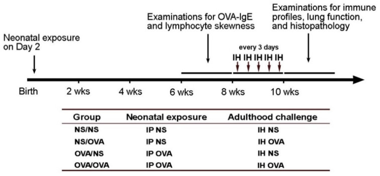
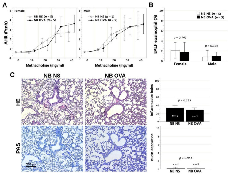
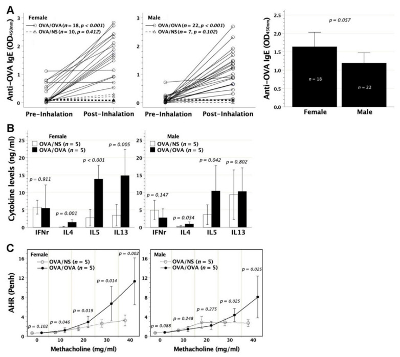
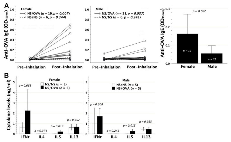
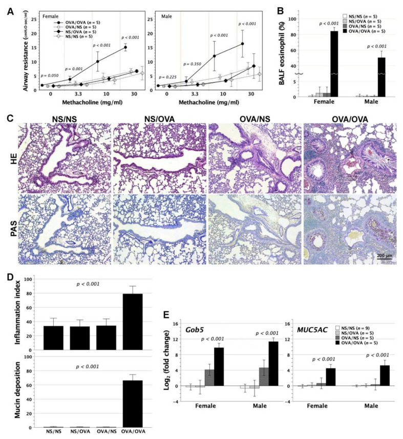
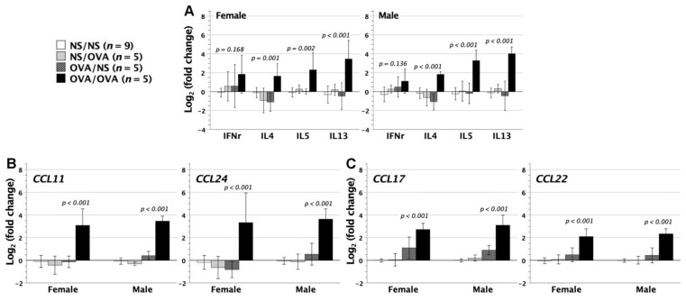
We previously demonstrated that fetal allergen exposure caused T-helper 2 (Th2) cell sensitization. Although neonates are immunologically more mature than fetuses, asthmatic lungs were reportedly mitigated by neonatal allergen administration, mechanically referring to regulatory T-cells and TGF-β signaling but lacking the immunological profiles after neonatal exposure. To reappraise the immunological outcome of neonatal allergen exposure, we injected adjuvant-free ovalbumin intraperitoneally into 2-day-old BALB/c neonates, followed by aerosolized ovalbumin inhalation in adulthood. Mice were examined for the immunological profiles specifically after neonatal exposures, lung function and histology (hematoxylin-eosin or periodic acid Schiff staining), and gene expressions of intrapulmonary cytokines (IL-4, IL-5, IL-13 and IFN-γ) and chemokines (CCL17, CCL22, CCL11 and CCL24). Neonatal ovalbumin exposure triggered Th2-skewed sensitization and ovalbumin-specific IgE production. Subsequent ovalbumin inhalation in adulthood boosted Th2 immunity and caused asthmatic lungs with structural and functional alterations of airways. Gender difference mainly involved airway hyperresponsiveness and resistance with greater female susceptibility to methacholine bronchospastic stimulation. In lungs, heightened chemoattractant gene expressions were only granted to neonatally ovalbumin-sensitized mice with aerosolized ovalbumin stress in adulthood, and paralleled by upregulated Th2 cytokine genes. Thus, aeroallergen stress in atopic individuals might upregulate the expression of intrapulmonary chemoattractants to recruit Th2 cells and eosinophils into the lungs, pathogenically linked to asthma development. Conclusively, murine neonates were sensitive to allergen exposures. Exposure events during neonatal stages were crucial to asthma predisposition in later life. These findings from a murine model point to allergen avoidance in neonatal life, possibly even very early in utero, as the best prospect of primary asthma prevention.

Keywords: adjuvant; asthma; chemokine; cytokine; neonate; ovalbumin; sensitization.


Conflict of interest statement

The authors declare no conflict of interest. The funders had no role in the design of the study; in the collection, analyses, or interpretation of data; in the writing of the manuscript, or in the decision to publish the results.

Figures

Figure 1
Figure 1
Schematic of experimental design for OVA exposure and challenge in mice, and grouping. IP, intraperitoneal injection; IH, inhalation; NS, normal saline; OVA, ovalbumin.
Figure 2
Figure 2
The generation of OVA-IgE and Th2 immune deviation following neonatal OVA exposure. BALB/c newborns (NB) were intraperitoneally exposed to adjuvant-free OVA of 50 μg at 2 days old (NB OVA). Serum OVA-IgE was measured by ELISA and lymphocyte skewness was in vitro examined under OVA stimulation. (A) At 6–8 weeks of age, both female and male recipients generated significant levels of OVA-IgE, as compared with control neonates receiving saline injection (NB NS). Serum OVA-IgE of NB OVA did not differ in mean titer between females and male (p = 0.076) (B) Lymphocyte polarization was examined by IFN-γ, IL-4, IL-5, and IL-13 secretions in cell cultures under OVA stimulation. NB OVA compared favorably in the levels of IL-4, IL-5, and IL-13 but not IFN-γ with NB NS, indicating a Th2-skewed phenotype. (C) Th2-skewed lymphocytes were characterized by IL-5 and IL-13 secretions at 7 days old, and (D) by IL-4, IL-5, and IL-13 secretions at 14 days old. Days 7 and 14 cytokine data were generated from subjects without specifying the gender due to the difficulty in visually telling female from male animals at this stage.
Figure 3
Figure 3
Pulmonary function and histology following neonatal OVA exposure. Following neonatal exposure to OVA of 50 μg (NB OVA), mice at 2 months old were subjected to pulmonary function test of AHR under aerosolized methacholine stimulation using whole-body plethysmograph. (A) In recipients regardless of gender, AHR expressed as Penh did not differ between NB OVA and saline controls (NB NS) upon bronchospastic stimuli at any aerosolized methacholine dose. (B) As to eosinophil percentage in BALF, no significant difference was observed between NB OVA and NB NS. (C) Representative images were taken from females with neonatal OVA exposure. There were no significant histological alterations in terms of leukocyte infiltration and goblet cell hyperplasia/mucin deposition, which were common to males. HE: hematoxylin and eosin; PAS: periodic acid Schiff.
Figure 4
Figure 4
Immunological outcome and airway hyperresponsiveness in neonatally OVA-sensitized mice following airway OVA stress. BALB/c neonates were intraperitoneally sensitized to OVA at 2 days old, and further subjected to repetitive OVA inhalation at 8–10 weeks old (OVA/OVA). (A) Both female and male recipients showed significantly raised levels of serum OVA-IgE after OVA inhalation (paired comparison), as opposed to neonatally OVA-sensitized mice subjected to saline inhalation (OVA/NS). The mean titer of serum OVA-IgE after OVA inhalation in OVA/OVA made no difference between females and males (p = 0.057). (B) In cell culture under OVA stimulation, group OVA/OVA exhibited enhanced lymphocyte capacity for generating Th2 cytokines of IL-4, IL-5, and IL-13 in females, but only IL-4 and IL-5 in males, as compared with group OVA/NS. It made no difference in the generation of Th1 IFN-γ between groups OVA/OVA and OVA/NS. (C) AHR significantly increased upon bronchospastic stimuli at 10–40 mg/mL of aerosolized methacholine in females, but at 30–40 mg/mL in males.
Figure 5
Figure 5
Immunological outcome of OVA inhalation in adulthood. Two-month-old BALB/c adults that had been given intraperitoneal saline injection at 2 days old were subjected to repetitive OVA inhalation every 3 days for a total of 5 times (NS/OVA). (A) OVA inhalation in both female and male recipients led to the secretion of serum OVA-IgE, as opposed to the recipients with neonatal saline injection followed by saline inhalation in adulthood (NS/NS). After OVA inhalation, the mean titer of serum OVA-IgE in NS/OVA did not differ between females and males (p = 0.062). (B) In lymphocyte culture under OVA stimulation, group NS/OVA regardless of gender compared favorably in the generation of IL-5 rather than IFN-γ, IL-4, and IL-13 with group NS/NS.
Figure 6
Figure 6
Airway resistance, histological alteration, and mucin-related gene expression in neonatally OVA-sensitized mice with aerosolized OVA stress in adulthood. Two-day-old BALB/c neonates underwent intraperitoneal OVA injection, and then received OVA inhalation 5 times at 2 months old (OVA/OVA). (A) Airway resistance to methacholine stimuli was measured in tracheostomized animals, using a FinePointeTM RC system. In group OVA/OVA, airway resistance notably rose in females as methacholine bronchospastic stimuli increased from 3.3 to 30 mg/mL, but in males from 10 to 30 mg/mL, as compared with that of groups OVA/NS, NS/NS, and NS/OVA. There was no significant difference in airway resistance among groups OVA/NS, NS/NS, and NS/OVA. (B) Group OVA/OVA compared favorably in BALF eosinophil percentage with groups OVA/NS, NS/NS and NS/OVA. (C) Histological examination revealed peribronchial and perivascular inflammation (HE staining), and extensive goblet cell hyperplasia of airways with luminal mucin deposition (PAS staining) in the lungs of group OVA/OVA. Representative images were taken from females. Females and males had common patterns of histological findings. (D) The severity of peribronchial/perivascular inflammation and mucin deposition on captured images was quantified by image analysis software. Group OVA/OVA compared favorably in integrated intensity of inflammation and mucin deposition with groups OVA/NS, NS/NS, and NS/OVA. (E) The expression of Gob5 and MUC5AC genes was determined by quantitative PCR and normalized to β-actin expression. The fold change was the expression ratio as the normalized expression level of a subject divided by the mean of normalized expression levels in group NS/NS. Group OVA/OVA compared favorably in Gob5 and MUC5AC expression with groups OVA/NS, NS/NS, and NS/OVA. There was no significant difference in Gob5 and MUC5AC expression among groups OVA/NS, NS/NS, and NS/OVA by post hoc multiple comparisons using Bonferroni method. OVA/NS: neonatal OVA exposure and adulthood saline inhalation; NS/NS: neonatal saline exposure and adulthood saline inhalation; NS/OVA: neonatal saline exposure and adulthood OVA inhalation.
Figure 7
Figure 7
Gene expression of cytokines and chemokines in the lungs. Neonatally OVA-sensitized mice with adulthood aerosolized OVA stress (OVA/OVA) were examined for cytokine and chemokine gene expression of the lungs. Control mice were neonatally saline-exposed mice with adulthood saline inhalation (NS/NS). (A) Regardless of gender, group OVA/OVA had significantly upregulated expression of IL-4, IL-5, and IL-13 for 22–4 folds as compared with group NS/NS, whereas IFN-γ gene expression did not differ between groups OVA/OVA and NS/NS. (B,C) Likewise, gene expression of chemokines CCL11, CCL24, CCL17, and CCL22 in the lungs showed 22–3-fold increase in group OVA/OVA, as opposed to group NS/NS. Neither group NS/OVA nor group OVA/NS had any significant changes of cytokines or chemokine gene expression when compared with group NS/NS.

Similar articles

Cited by

References

    1. Billingham R.E., Brent L., Medawar P.B. ‘Actively Acquired Tolerance’ of Foreign Cells. Nat. Cell Biol. 1953;172:603–606. doi: 10.1038/172603a0. - DOI - PubMed
    1. Chen J.-C., Chan C.-C., Wu C.-J., Ou L.-S., Yu H.-Y., Chang H.-L., Tseng L.-Y., Kuo M.-L. Fetal Phagocytes Take up Allergens to Initiate T-Helper Cell Type 2 Immunity and Facilitate Allergic Airway Responses. Am. J. Respir. Crit. Care Med. 2016;194:934–947. doi: 10.1164/rccm.201508-1703OC. - DOI - PubMed
    1. Chen J.-C., Kuo M.-L., Ou L.-S., Chang P.-Y., Muench M.O., Shen C.-R., Chang H.-L., Yu H.-Y., Fu R.-H. Characterization of Tolerance Induction through Prenatal Marrow Transplantation: The Requirement for a Threshold Level of Chimerism to Establish Rather than Maintain Postnatal Skin Tolerance. Cell Transplant. 2010;19:1609–1622. doi: 10.3727/096368910X516583. - DOI - PubMed
    1. Chen J.-C. Immunological Consequences of In Utero Exposure to Foreign Antigens. Front. Immunol. 2021;12:638435. doi: 10.3389/fimmu.2021.638435. - DOI - PMC - PubMed
    1. Smith R.T., Bridges R.A., Thomson E. Immunological unresponsiveness in rabbits produced by neonatal injection of defined antigens. J. Exp. Med. 1958;108:227–250. doi: 10.1084/jem.108.2.227. - DOI - PMC - PubMed

LinkOut - more resources