. 2021 Jun 9;22(12):6216.
doi: 10.3390/ijms22126216.

Altered Expression of ESR1, ESR2, PELP1 and c-SRC Genes Is Associated with Ovarian Cancer Manifestation

Affiliations

Altered Expression of ESR1, ESR2, PELP1 and c-SRC Genes Is Associated with Ovarian Cancer Manifestation

Monika Englert-Golon et al. Int J Mol Sci. .

Abstract

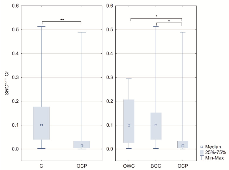
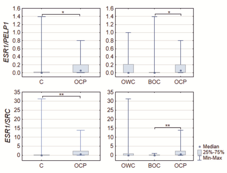
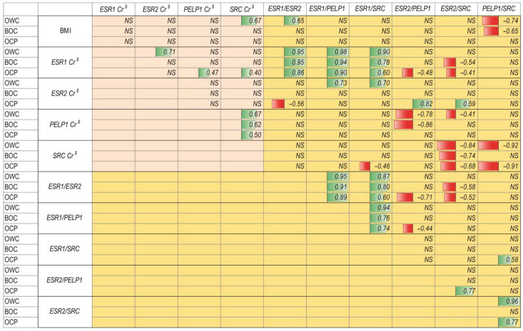
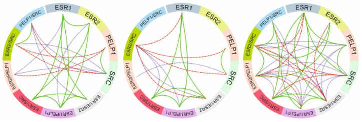
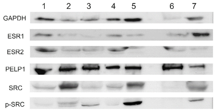
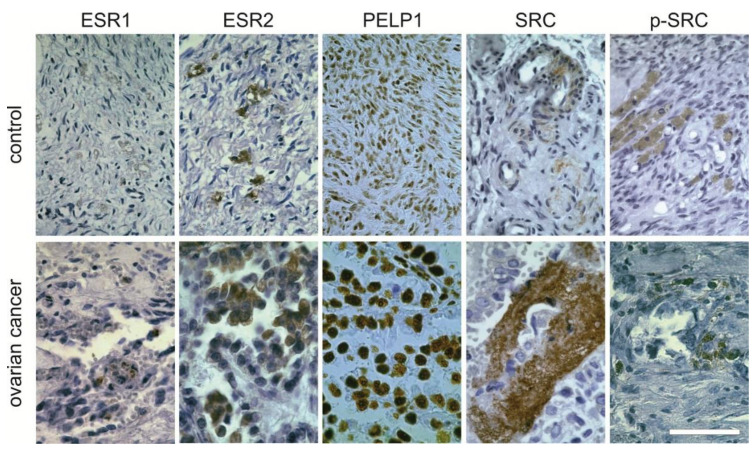
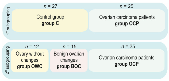
Ovarian cancer remains the leading cause of death due to gynecologic malignancy. Estrogen-related pathways genes, such as estrogen receptors (ESR1 and ESR2) and their coregulators, proline-, glutamic acid-, and leucine-rich protein 1 (PELP1), and proto-oncogene tyrosine-protein kinase c-Src (SRC) are involved in ovarian cancer induction and development, still they require in-depth study. In our study, tissue samples were obtained from 52 females of Caucasian descent (control group without cancerous evidence (n = 27), including noncancerous benign changes (n = 15), and the ovarian carcinoma (n = 25)). Using quantitative analyses, we investigated ESRs, PELP1, and SRC mRNA expression association with ovarian tumorigenesis. Proteins' presence and their location were determined by Western blot and immunohistochemistry. Results showed that PELP1 and SRC expression levels were found to differ in tissues of different sample types. The expression patterns were complex and differed in the case of ovarian cancer patients compared to controls. The most robust protein immunoreactivity was observed for PELP1 and the weakest for ESR1. The expression patterns of analyzed genes represent a potentially interesting target in ovarian cancer biology, especially PELP1. This study suggests that specific estrogen-mediated functions in the ovary and ovary-derived cancer might result from different local interactions of estrogen with their receptors and coregulators.

Keywords: estrogen receptors (ESR1 and ESR2); estrogen signal transduction coregulators; ovarian cancer; proline-, glutamic acid-, and leucine-rich protein 1 (PELP1); proto-oncogene tyrosine-protein kinase c-Src (SRC).


Conflict of interest statement

The authors declare no conflict of interest.

Figures

Figure 1
Figure 1
Boxplots of the SRC normalized concentration ratios in controls (C), ovarian cancer patients (OCP), ovarian tissue samples lack of any changes patients (OWC) and with benign noncancerous changes (BOC); * p < 0.05 and ** p < 0.01. Medians, interquartile range and p-values are shown in Table 1.
Figure 2
Figure 2
Boxplots of the ESR1/PELP1 and ESR1/SRC quotient ratios in controls (C), ovarian cancer patients (OCP), ovarian tissue samples lack of any changes patients (OWC) and with benign noncancerous changes (BOC) * p < 0.05 and ** p < 0.01. Medians, interquartile range and p-values are shown in Table 1.
Figure 3
Figure 3
Correlation coefficient plot of ovarian tissue samples lack of any changes patients (OWC) females with benign noncancerous changes in the ovary (BOC) and ovarian cancer patients. Designation: NS—not significant; numbers—Spearman rank correlation coefficients; green bars—proportional correlation; red bars—reverse proportional correlation. Green background—clinical data correlations; pink background—gene expression correlations; yellow background—gene expression quotient ratio correlations. Cr $ concentration ratios data normalized using min-max normalization.
Figure 4
Figure 4
Circular correlation coefficient plot of gene expression and expression quotient. Reverse proportional correlations were indicated by red dash lines, proportional correlation by solid green lines and violet dotted lines indicated significant correlation coefficients relationship in gene-to-gene expression and quotient-to-quotient influence. Ovary without changes (left), benign noncancerous tissue samples (middle), and ovarian cancer (right).
Figure 5
Figure 5
Western blot analysis in selected cases of ovarian cancer tissues (1–5) and noncancerous ovary (6–7).
Figure 6
Figure 6
Immunohistochemical staining for analyzed proteins in controls and ovarian cancer tissue. Positive staining is shown in brown. The bar represents 100 µm. This section may be divided by subheadings. It should provide a concise and precise description of the experimental results, their interpretation, as well as the experimental conclusions that can be drawn.
Figure 7
Figure 7
Study material. Diagram showing the number of tissue samples, allocated to different examined groups.
Figure 8
Figure 8
Amplicons of the analyzed genes ESR1, PELP1 and SRC with their primers, TaqMan® Probes and introns positions. ESR2 and HPRT (reference) probe assays are protected by trade secrets.

References

    1. Reid B.M., Permuth J.B., Sellers T.A. Epidemiology of ovarian cancer: A review. Cancer Biol. Med. 2017;14:9–32. - PMC - PubMed
    1. Boussios S., Karihtala P., Moschetta M., Abson C., Karathanasi A., Zakynthinakis-Kyriakou N., Ryan J.E., Sheriff M., Rassy E., Pavlidis N. Veliparib in ovarian cancer: A new synthetically lethal therapeutic approach. Investig. New Drugs. 2020;38:181–193. doi: 10.1007/s10637-019-00867-4. - DOI - PubMed
    1. Dochez V., Caillon H., Vaucel E., Dimet J., Winer N., Ducarme G. Biomarkers and algorithms for diagnosis of ovarian cancer: CA125, HE4, RMI and ROMA, a review. J. Ovarian Res. 2019;12:28. doi: 10.1186/s13048-019-0503-7. - DOI - PMC - PubMed
    1. Jelovac D., Armstrong D.K. Recent progress in the diagnosis and treatment of ovarian cancer. CA Cancer J. Clin. 2011;61:183–203. doi: 10.3322/caac.20113. - DOI - PMC - PubMed
    1. Modugno F., Laskey R., Smith A.L., Andersen C.L., Haluska P., Oesterreich S. Hormone response in ovarian cancer: Time to reconsider as a clinical target? Endocr. Relat. Cancer. 2012;19:R255–R279. doi: 10.1530/ERC-12-0175. - DOI - PMC - PubMed

MeSH terms