

Imaging of Substantia Nigra in Parkinson's Disease: A Narrative Review
- PMID: 34207681
- PMCID: PMC8230134
- DOI: 10.3390/brainsci11060769
Imaging of Substantia Nigra in Parkinson's Disease: A Narrative Review
Abstract
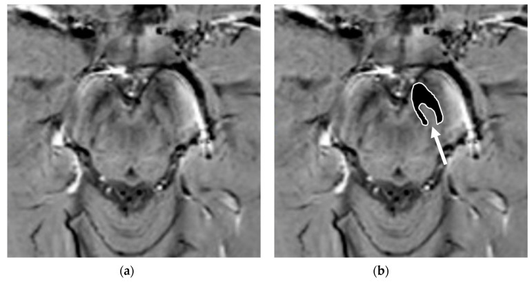
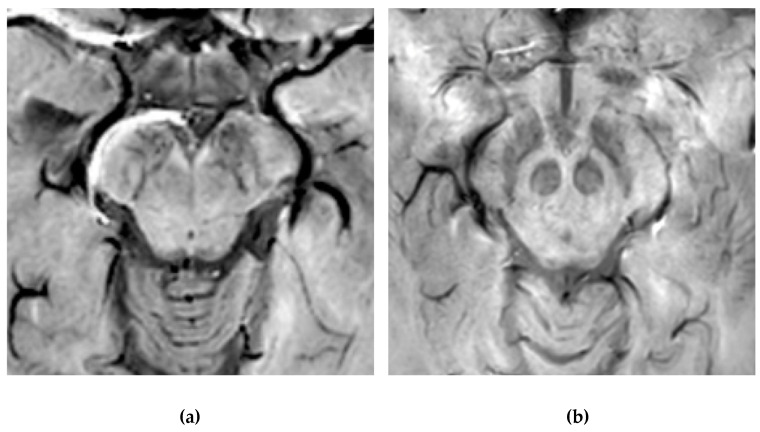
Parkinson's disease (PD) is a progressive neurodegenerative disorder, characterized by motor and non-motor symptoms due to the degeneration of the pars compacta of the substantia nigra (SNc) with dopaminergic denervation of the striatum. Although the diagnosis of PD is principally based on a clinical assessment, great efforts have been expended over the past two decades to evaluate reliable biomarkers for PD. Among these biomarkers, magnetic resonance imaging (MRI)-based biomarkers may play a key role. Conventional MRI sequences are considered by many in the field to have low sensitivity, while advanced pulse sequences and ultra-high-field MRI techniques have brought many advantages, particularly regarding the study of brainstem and subcortical structures. Nowadays, nigrosome imaging, neuromelanine-sensitive sequences, iron-sensitive sequences, and advanced diffusion weighted imaging techniques afford new insights to the non-invasive study of the SNc. The use of these imaging methods, alone or in combination, may also help to discriminate PD patients from control patients, in addition to discriminating atypical parkinsonian syndromes (PS). A total of 92 articles were identified from an extensive review of the literature on PubMed in order to ascertain the-state-of-the-art of MRI techniques, as applied to the study of SNc in PD patients, as well as their potential future applications as imaging biomarkers of disease. Whilst none of these MRI-imaging biomarkers could be successfully validated for routine clinical practice, in achieving high levels of accuracy and reproducibility in the diagnosis of PD, a multimodal MRI-PD protocol may assist neuroradiologists and clinicians in the early and differential diagnosis of a wide spectrum of neurodegenerative disorders.
Keywords: Parkinson’s disease; biomarkers; iron; magnetic resonance imaging; neurodegenerative diseases; neuromelanin; nigrosome-1; parkinsonian disorders; radiomics.
Conflict of interest statement
The authors declare no conflict of interest.
Figures


References
Publication types
LinkOut - more resources
Full Text Sources
Medical
Miscellaneous
