. 2021 Jun 9;22(12):6241.
doi: 10.3390/ijms22126241.

Decellularized Porcine Cartilage Scaffold; Validation of Decellularization and Evaluation of Biomarkers of Chondrogenesis

Affiliations

Decellularized Porcine Cartilage Scaffold; Validation of Decellularization and Evaluation of Biomarkers of Chondrogenesis

Roxanne N Stone et al. Int J Mol Sci. .

Abstract

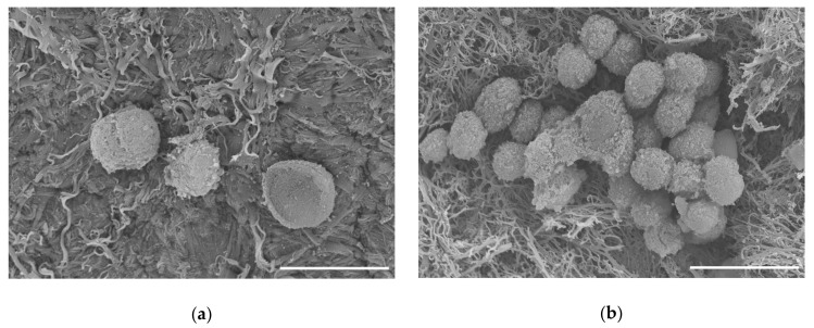
Osteoarthritis is a major concern in the United States and worldwide. Current non-surgical and surgical approaches alleviate pain but show little evidence of cartilage restoration. Cell-based treatments may hold promise for the regeneration of hyaline cartilage-like tissue at the site of injury or wear. Cell-cell and cell-matrix interactions have been shown to drive cell differentiation pathways. Biomaterials for clinically relevant applications can be generated from decellularized porcine auricular cartilage. This material may represent a suitable scaffold on which to seed and grow chondrocytes to create new cartilage. In this study, we used decellularization techniques to create an extracellular matrix scaffold that supports chondrocyte cell attachment and growth in tissue culture conditions. Results presented here evaluate the decellularization process histologically and molecularly. We identified new and novel biomarker profiles that may aid future cartilage decellularization efforts. Additionally, the resulting scaffold was characterized using scanning electron microscopy, fluorescence microscopy, and proteomics. Cellular response to the decellularized scaffold was evaluated by quantitative real-time PCR for gene expression analysis.

Keywords: C28/I2 cells; cartilage; chondrocytes; decellularized; histology; porcine auricular cartilage; proteomics; real time quantitative PCR; scaffold; scanning electron microscopy.


Conflict of interest statement

The authors declare no conflict of interest. The funders had no role in the design of the study; in the collection, analyses, or interpretation of data; in the writing of the manuscript; or in the decision to publish the results.

Figures

Figure 1
Figure 1
Cartilage scaffold resulting from decellularization process: (a) porcine auricular cartilage after dissection and initial processing; (b) 8 mm diameter cartilage disc after decellularization process was complete. Discs of decellularized cartilage were formed using an 8 mm diameter biopsy punch. Scale bars: (a) 30 mm; (b) 2 mm.
Figure 2
Figure 2
Flowchart for decellularization process.
Figure 3
Figure 3
Visualization of cartilage tissue by histology. (a) Nondecellularized porcine cartilage stained with H&E, 10× transverse image. (b) H&E stain on final decellularized cartilage, transverse image. (c) Hoechst stain to fluorescently visualize DNA (blue). Arrows indicate nuclei. (d) Hoechst stain shows absence of DNA after decellularization process (see absence of blue). Scale bar = 100 µm for (a,b); scale bar = 50 µm for (c,d).
Figure 4
Figure 4
DNA content before, during sequential stages, and after decellularization process. DNA was extracted and quantified spectrophotometrically. Quantitative measurements of DNA within scaffolds before, during, and after decellularization process indicated that residual DNA was at approximately 24.76%, 2.63%, 0.66%, and finally 0.22% of the original content. Error bars: Mean ± standard error of the mean. N = 6.
Figure 5
Figure 5
Visualization of cartilage by scanning electron microscopy. (a) Cartilage prior to treatment; (b) higher magnification of cartilage prior to treatment, where the tissue appears smooth and intact; (c) final decellularized cartilage disc; (d) higher magnification of decellularized cartilage disc. Appearance after decellularization indicates a rough surface with increased surface area and exposed collagen fibrillar networks. Scale bars = 20 µm.
Figure 6
Figure 6
Collagenous composition of the decellularized scaffold. Blue bars show the collagens present in cartilage prior to the decellularization process, and red bars show composition of the scaffold after the decellularization process. The length of the bars to the left and right corresponds to abundance of the specific collagens. Collagen alpha chains are listed in the order of decreasing prevalence within native cartilage. The resulting decellularized scaffold (red) contained COL2A1, COL1A1, COL6A3, COL1A2, COL11A2, COL11A1, COL4A5, COL3A1, COL5A2, COL16A1, COL5A1, COL4A2, COL4A3, COL5A3, COL27A1, COL13A1, COL4A1, COL12A1, and COL17A1. Minor contributions of COL7A1, COL22A1, COL28A1, COL4A4, COL9A2, COL6A2, COL14A1, COL8A2, COL18A1, COL23A1, COL6A6, and COL20A1 were detected after decellularization. Horizontal axes values represent the sum of peptide spectrum matches (PSM), the total number of identified peptides for each collagen as an indication of quantity.
Figure 7
Figure 7
Scanning electron micrographs of C28/I2 chondrocyte cells on decellularized scaffold. (a) Cells attached to scaffold after 1 week in culture. (b) Cells on scaffold after 8 months in culture. Scale bar = 20 µm.
Figure 8
Figure 8
Analysis of variance of difference to determine most suitable housekeeping genes. Candidate housekeeping genes were considered in a pairwise fashion to determine which ones were the most consistently expressed independent of experimental condition within this study. GAPDH and HPRT1 displayed the minimum variance of difference over all experimental conditions and timepoints.
Figure 9
Figure 9
Correlation analysis of gene expression during growth on tissue culture plastic and cartilage scaffold. C28/I2 cells were grown under conventional conditions on tissue culture plastic for one week (a) and compared to cells grown on cartilage scaffold for 7 days (b). The diagonal line in (a,b) indicates the trend expected if there was no change between conditions; day 0 versus day 7 on tissue culture plastic in (a) and day 7 on tissue culture plastic versus day 7 on cartilage scaffold in (b). Data points above the line reflect genes expressed at higher levels on the cartilage scaffold compared to plastic. Data points below the line indicate genes that were expressed at higher levels on plastic compared to the cartilage scaffold. Data points that fall on the line were not changed.
Figure 10
Figure 10
Gene expression of markers in C28/I2 cells. Gene expression for extracellular matrix, cell adhesion molecules, and cell–cell attachment proteins was measured by quantitative real-time PCR. The venn diagram clusters genes with respect to upregulation in cells grown on conventional tissue culture plastic shown in yellow, in cells grown on the decellularized cartilage scaffold shown in pink. Genes that were upregulated under both conditions are shown in the orange overlap region of the Venn diagram.

References

    1. Cross M., Smith E., Hoy D., Nolte S., Ackerman I., Fransen M., Bridgett L., Williams S., Guillemin F., Hill C.L., et al. The global burden of hip and knee osteoarthritis: Estimates from the Global Burden of Disease 2010 study. Ann. Rheum. Dis. 2014;73:1323–1330. doi: 10.1136/annrheumdis-2013-204763. - DOI - PubMed
    1. Escobar Ivirico J.L., Bhattacharjee M., Kuyinu E., Nair L.S., Laurencin C.T. Regenerative Engineering for Knee Osteoarthritis Treatment: Biomaterials and Cell-Based Technologies. Engineering. 2017;3:16–27. doi: 10.1016/J.ENG.2017.01.003. - DOI - PMC - PubMed
    1. Helmick C.G., Felson D.T., Lawrence R.C., Gabriel S., Hirsch R., Kwoh C.K., Liang M.H., Maradit Kremers H., Mayes M.D., Merkel P.A., et al. National Arthritis Data Workgroup Estimates of the prevalence of arthritis and other rheumatic conditions in the United States: Part I. Arthritis Rheum. 2008;58:15–25. doi: 10.1002/art.23177. - DOI - PubMed
    1. Toh W.S., Foldager C.B., Pei M., Hui J.H.P. Advances in Mesenchymal Stem Cell-based Strategies for Cartilage Repair and Regeneration. Stem Cell Rev. Rep. 2014;10:686–696. doi: 10.1007/s12015-014-9526-z. - DOI - PubMed
    1. Gupta P.K., Das A.K., Chullikana A., Majumdar A.S. Mesenchymal stem cells for cartilage repair in osteoarthritis. Stem Cell Res. Ther. 2012;3:25. doi: 10.1186/scrt116. - DOI - PMC - PubMed

Publication types

LinkOut - more resources