. 2021 Jun 11;9(6):1270.
doi: 10.3390/microorganisms9061270.

Malnutrition Aggravates Alterations Observed in the Gut Structure and Immune Response of Mice Infected with Leishmania infantum

Affiliations

Malnutrition Aggravates Alterations Observed in the Gut Structure and Immune Response of Mice Infected with Leishmania infantum

Felipe Gaitán-Albarracín et al. Microorganisms. .

Abstract

Malnutrition is a risk factor for developing visceral leishmaniasis and its severe forms. Our group demonstrated that malnourished animals infected with Leishmania infantum had severe atrophies in lymphoid organs and T cell subpopulations as well as altered levels of thymic and splenic chemotactic factors, all of which resulted in dysfunctional lymphoid microenvironments that promoted parasite proliferation. Here, we hypothesize that malnutrition preceding parasite infection leads to structural and immunological changes in the gut mucosae, resulting in a failure in the immune response sensed in the intestine. To evaluate this, we analyzed the immunopathological events resulting from protein malnutrition in the guts of BALB/c mice infected with L. infantum. We observed lymphocytic/lymphoplasmacytic inflammatory infiltrates and lymphoid hyperplasia in the duodenum of well-nourished-infected mice; such alterations were worsened when malnutrition preceded infection. Parasite infection induced a significant increase of duodenal immunoglobulin A (IgA) of well-nourished animals, but those levels were significantly decreased in malnourished-infected mice. In addition, increased levels of Th17-related cytokines in duodenums of malnourished animals supported local inflammation. Together, our results suggest that the gut plays a potential role in responses to L. infantum infection-and that such responses are impaired in malnourished individuals.

Keywords: IgA; Leishmania infantum; duodenum; gut; inflammation; malnutrition; visceral leishmaniasis.


Conflict of interest statement

The authors declare no conflict of interest. The funders had no role in the design of the study; in the collection, analyses, or interpretation of data; in the writing of the manuscript, or in the decision to publish the results.

Figures
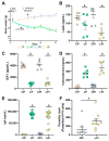
Figure 1
Figure 1
Bodyweight and hormonal levels in malnourished BALB/c mice infected with L. infantum. (A) Male BALB/c mice were fed a 14% (n = 24, control protein (CP)) or 4% (n = 24, low protein (LP)) protein diet for 21 days. On day 7 of the experimental period, half of the animals were infected with L. infantum and the other half received an injection of saline solution. Bodyweight was recorded every third day and expressed as average ± SEM; n = 12 mice in each group. Statistical differences before the day of infection were determined by Student’s t-test (p < 0.0005). After infection, a two-way ANOVA with Tukey’s post-hoc test was used (p < 0.0001). (B) Leptin, (C) total IGF-1 (Insulin-like growth factor 1), (D) corticosterone, and (E) immunoglobulin A (IgA) serum levels were analyzed in the experimental groups (n = 6 per group). Leptin, IGF-1, and IgA were measured by commercial ELISA assays, whereas corticosterone was measured by radioimmunoassay. Statistical differences due to diet (a), infection (b), or (c) interaction between diet and infection were determined by two-way ANOVA (p < 0.05). (F) Parasite load was determined by qPCR in the spleen and statistical differences were determined by Student’s t-test (* p < 0.05). CP: control protein, animals fed 14% protein diet; LP: low protein, animals fed 4% protein diet, CPi: animals fed 14% protein diet and infected; LPi: animals fed 4% protein diet and infected.
Figure 2
Figure 2
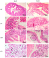
Histopathological analysis of duodenum and jejunum from BALB/c mice. CP: animals fed 14% protein diet, LP: animals fed 4% protein diet, CPi: animals fed 14% protein diet and infected, LPi: animals fed 4% protein diet and infected. (AD) Duodenum. H&E. (A) CP mouse without histological alterations. (B) LP mouse showing mild and focal lymphocytic infiltrate (circle and inset) in the lamina propria. (C) CPi mouse showing moderate to severe and diffuse lymphocytic infiltrate in the lamina propria. (D) LPi mouse showing moderate to severe and diffuse lymphoplasmacytic infiltrate (circle and inset). (EH) Jejunum. H&E (E) CP mouse without histological alterations. (F) LP mouse showing mild and focal lymphocytic infiltrate (circle and inset) in the lamina propria. (G) CPi mouse without histological alterations. (H) LPi mouse showing moderate to severe lymphoplasmacytic infiltrate (circle and inset) in the lamina propria.
Figure 3
Figure 3
Histopathological analysis of ileum and colon from BALB/c mice. CP: animals fed 14% protein diet, LP: animals fed 4% protein diet, CPi: animals fed 14% protein diet and infected, LPi: animals fed 4% protein diet and infected. (A-D) Ileum. H&E. (A) CP mouse without histological alterations. (B) LP mouse showing mild and diffuse lymphocytic infiltrate (circle and inset) in the lamina propria. (C) CPi mouse showing mild and diffuse lymphoplasmacytic infiltrate (circle and inset) in the lamina propria. (D) LPi mouse showing moderate to severe and diffuse lymphoplasmacytic infiltrate (circle and inset) in the lamina propria. (EH) Colon. H&E (E) CP mouse without histological alterations. (F) LP mouse showing moderate to severe and diffuse lymphocytic infiltrate in the lamina propria. (G) CPi mouse showing mild and diffuse lymphocytic infiltrate (circle and inset) in the lamina propria. (H) LPi mouse showing mild and diffuse lymphoplasmacytic infiltrate (circle and inset) in the lamina propria.
Figure 4
Figure 4
Total score of histopathological changes observed in the intestine regions of malnourished BALB/c mice infected with L. infantum. (A) The score values represent the sum of the changes observed in each experimental group according to the presence or absence of the alteration and its degree of severity (Table S2). (B) Total score of immunological and structural changes observed in each experimental group. n = 6 animals per group. CP: animals fed 14% protein diet, LP: animals fed 4% protein diet, CPi: animals fed 14% protein diet and infected, LPi: animals fed 4% protein diet and infected.
Figure 5
Figure 5
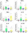
Cytokine expression in the duodenum from malnourished BALB/c mice infected with L. infantum. il-12, ifnγ, tnfα, tgfβ and il-17 mRNA expression levels measured by qPCR in the duodenum of each experimental group. The values are expressed as normalized ratios between the target gene expression and the geometric median of the ATP-5, GAPDH and CYC-1 genes. Statistical differences due to (a) diet (p < 0.001) were determined by two-way ANOVA. CP: animals fed 14% protein diet; LP: animals fed 4% protein diet, CPi: animals fed 14% protein diet and infected; LPi: animals fed 4% protein diet and infected.
Figure 6
Figure 6
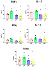
Cytokines levels in the luminal fluid of duodenum from malnourished BALB/c mice infected with L. infantum. TNFα, IL-12, IFNγ, IL-13 and TGFβ protein levels were measured in the luminal fluid of the duodenum at 14 days post-infection using a flow cytometry-based multiplex assay. Significant differences due to infection (b), p < 0.01 for TNFα and IL-12 and p < 0.05 for TGFβ, as determined by two-way ANOVA. CP: animals fed 14% protein diet; LP: animals fed 4% protein diet, CPi: animals fed 14% protein diet and infected; LPi: animals fed 4% protein diet and infected.
Figure 7
Figure 7
Protein levels of Th-17 related cytokines in the luminal fluid of duodena from malnourished BALB/c mice infected with L. infantum. IL-17A, IL-21, IL-22, and IL-23 protein levels in the luminal fluid of duodena were measured at 14 days post-infection using a flow cytometry-based multiplex assay. a: significant differences due to diet (p < 0.05) determined by two-way ANOVA. CP: animals fed 14% protein diet; LP: animals fed 4% protein diet, CPi: animals fed 14% protein diet and infected; LPi: animals fed 4% protein diet and infected.
Figure 8
Figure 8
Chemokine levels in the luminal fluid of duodenum from malnourished BALB/c mice infected with L. infantum. CCL5, CXCL9, and CXCL10 levels in the luminal fluid of duodena were measured at 14 days post-infection using a flow cytometry-based multiplex assay. Significant differences due to diet (a), p < 0.05; significant differences due to infection (b), p < 0.01; significant differences due to interaction between diet and infection (c), p < 0.05, as determined by two-way ANOVA. CP: animals fed 14% protein diet; LP: animals fed 4% protein diet; CPi: animals fed 14% protein diet and infected; LPi: animals fed 4% protein diet and infected.
Figure 9
Figure 9
Levels of secreted IgA in duodenum of malnourished BALB/c mice infected with L. infantum. The levels of secreted IgA measured by ELISA according to the protocol described in materials and methods. Significant differences due to infection (b), p < 0.05; significant differences due to interaction between diet and infection (c), p < 0.01. CP: animals fed 14% protein diet; LP: animals fed 4% protein diet, CPi: animals fed 14% protein diet and infected; LPi: animals fed 4% protein diet and infected.

Similar articles

Cited by

References

    1. Bourke C.D., Berkley J.A., Prendergast A.J. Immune Dysfunction as a Cause and Consequence of Malnutrition. Trends Immunol. 2016;37:386–398. doi: 10.1016/j.it.2016.04.003. - DOI - PMC - PubMed
    1. Badaro R., Carvalho E.M., Rocha H., Queiroz A.C., Jones T.C. Leishmania donovani: An opportunistic microbe associated with progressive disease in three immunocompromised patients. Lancet. 1986;1:647–649. doi: 10.1016/S0140-6736(86)91725-3. - DOI - PubMed
    1. Rosenthal P.J., Chaisson R.E., Hadley W.K., Leech J.H. Rectal leishmaniasis in a patient with acquired immunodeficiency syndrome. Am. J. Med. 1988;84:307–309. doi: 10.1016/0002-9343(88)90430-5. - DOI - PubMed
    1. Anstead G.M., Chandrasekar B., Zhang Q., Melby P.C. Multinutrient undernutrition dysregulates the resident macrophage proinflammatory cytokine network, nuclear factor-kappaB activation, and nitric oxide production. J. Leukoc. Biol. 2003;74:982–991. doi: 10.1189/jlb.0203064. - DOI - PubMed
    1. Anstead G.M., Chandrasekar B., Zhao W., Yang J., Perez L.E., Melby P.C. Malnutrition alters the innate immune response and increases early visceralization following Leishmania donovani infection. Infect. Immun. 2001;69:4709–4718. doi: 10.1128/IAI.69.8.4709-4718.2001. - DOI - PMC - PubMed

LinkOut - more resources