Comparative Study
. 2021 Jun 11;19(6):334.
doi: 10.3390/md19060334.

A Comparative In Vitro Evaluation of the Anti-Inflammatory Effects of a Tisochrysis lutea Extract and Fucoxanthin

Affiliations
Comparative Study

A Comparative In Vitro Evaluation of the Anti-Inflammatory Effects of a Tisochrysis lutea Extract and Fucoxanthin

Elisabetta Bigagli et al. Mar Drugs. .

Abstract

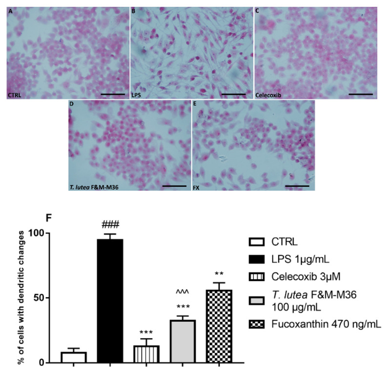
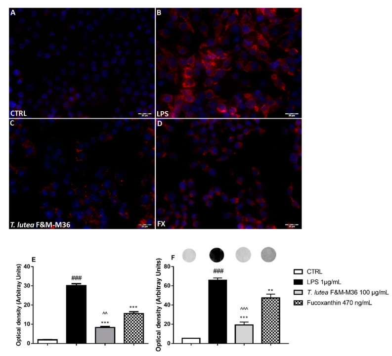
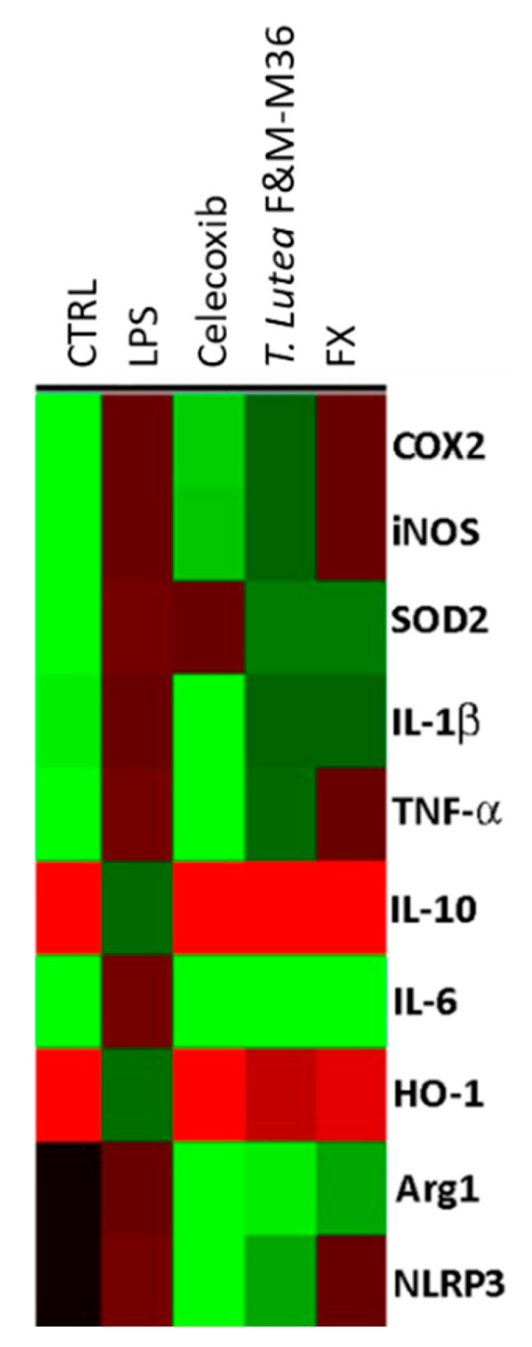
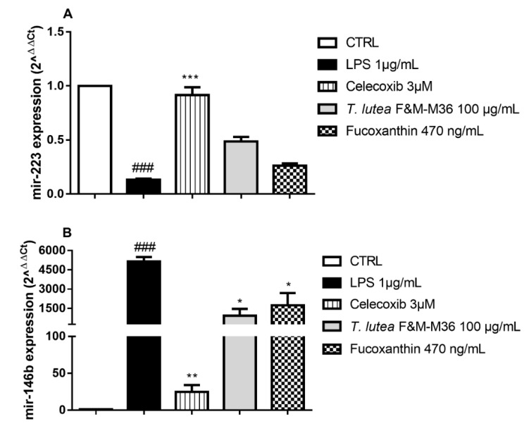
In this study, we compared the effects of a Tisochrysis lutea&nbsp;(T. lutea) F&M-M36 methanolic extract with those of fucoxanthin (FX) at equivalent concentration, on lipopolysaccharide (LPS)-stimulated RAW 264.7 macrophages. The T. lutea F&M-M36 methanolic extract contained 4.7 mg of FX and 6.22 mg of gallic acid equivalents of phenols per gram. HPLC analysis revealed the presence of simple phenolic acid derivatives. The T. lutea F&M-M36 extract exhibited a potent and concentration-dependent inhibitory activity against COX-2 dependent PGE2 production compared to FX alone. Compared to LPS, T. lutea F&M-M36 extract and FX reduced the expression of IL-6 and of Arg1 and enhanced that of IL-10 and of HO-1; T. lutea F&M-M36 extract also significantly abated the expression of NLRP3, enhanced mir-223 expression and reduced that of mir-146b, compared to LPS (p < 0.05). These findings indicate that T. lutea F&M-M36 methanolic extract has a peculiar anti-inflammatory activity against COX-2/PGE2 and NLRP3/mir-223 that might be attributable to the known anti-inflammatory effects of simple phenolic compounds found in the extract that may synergize with FX. Our data suggest that T. lutea F&M-M36 may serve as a source of anti-inflammatory compounds to be further evaluated in in vivo models of inflammation.

Keywords: RAW 264.7; Tisochrysis lutea; fucoxanthin; inflammation; microRNA; microalgae.


Conflict of interest statement

T. lutea F&M-M36 belongs to the Culture Collection F&M S.r.l. culture collection, where M.R.T. and L.R. have a financial interest. The other authors have no conflicts of interest.

Figures

Figure 1
Figure 1
Chromatograms obtained by high-performance liquid chromatography coupled to a diode array detector—HPLC-DAD (280 nm) of methanolic extract of T. lutea F&M-M36. Peak 1–13: phenolic acid derivatives; pick 14: catechin derivative. The putative identification was conducted based on UV-vis absorption and retention time, in comparison with standards and literature data.
Figure 2
Figure 2
Morphology images of RAW 264.7 cells in different groups obtained by a light microscope. Hematoxylin and eosin staining of cells from different experimental groups: (A) Unstimulated RAW264.7 cells; (B) LPS-stimulated RAW264.7 cells; (C) LPS-stimulated RAW264.7 cells treated with Celecoxib 3 µM; (D) LPS-stimulated RAW264.7 cells treated with T. lutea F&M-M36 extract 100 µg/mL; (E) LPS-stimulated RAW264.7 cells treated with FX 470 ng/mL; (F) Percentage of cells with dendritic changes. ### p < 0.001 vs. unstimulated RAW 264.7 macrophages (CTRL); ** p < 0.01 and *** p < 0.001 vs. LPS ^^^ p < 0.001 vs. FX by ANOVA test and Dunnett’s Multiple Comparison Test. Data are expressed as mean ± SEM of five replicates. Magnification = 400×; Scale bar = 20 μm.
Figure 3
Figure 3
Effect of T. lutea F&M-M36 extract and FX on PGE2 production in RAW 264.7 stimulated with LPS for 18 h. ### p < 0.001 vs. unstimulated RAW 264.7 macrophages (CTRL); *** p < 0.001 vs. LPS ^^^ p < 0.001 vs. FX by ANOVA test and Dunnett’s Multiple Comparison test. Data are expressed as the mean ± SEM of four replicates.
Figure 4
Figure 4
Effect of T. lutea F&M-M36 extract and FX on COX-2 protein expression in LPS-stimulated RAW 264.7 cells. Panels (AD): COX-2 protein expression determined by immunocytochemistry with an anti-COX-2 antibody (red fluorescence). Nuclei were counterstained with DAPI (blue fluorescence); Magnification = 400×; Scale bar = 20 μm. Panel (E): Densitometric analysis of cells positive for COX-2. Panel (F): Densitometric analysis of dot blot results on COX-2 protein expression; above bars, representative dot blot images are shown. ### p < 0.001 vs. unstimulated RAW 264.7 macrophages (CTRL). ** p < 0.01 and *** p < 0.001 vs. LPS. ^^ p < 0.01 and ^^^ p < 0.001 vs. FX by ANOVA test and Dunnett’s Multiple Comparison test. Data are expressed as means ± SEM of four replicates.
Figure 5
Figure 5
Gene expression profiles of unstimulated RAW 264.7 macrophages (CTRL), RAW 264.7 macrophages stimulated with LPS and those treated with LPS in the presence of T. lutea F&M-M36 extract at 100 µg/mL and FX at 470 ng/mL. Each column represents a different treatment and each row a different gene; the color code indicates down-regulation (green) or up-regulation (red) compared to LPS.
Figure 6
Figure 6
Effect of T. lutea F&M-M36 extract and FX on mir-223 (Panel A) and mir-146b (Panel B) expression in LPS-stimulated RAW 264.7 cells. ### p < 0.001 vs. CTRL; * p < 0.05, ** p < 0.01 and *** p < 0.001 vs. LPS by one-way ANOVA and Dunnett’s Multiple Comparisons test.

References

    1. Bendif E., Probert I., Schroeder D.C., de Vargas C. On the description of Tisochrysis lutea gen. nov., sp. nov. and Isochrysis nuda sp. nov. in the Isochrysidales, and the transfer of Dicrateria to the Prymnesiales (Haptophyta) J. Appl. Phycol. 2013;25:1763–1776. doi: 10.1007/s10811-013-0037-0. - DOI
    1. Camacho-Rodríguez J., Cerón-García M.C., González-López C.V., López-Rosales L., Contreras-Gómez A., Molina-Grima E. Use of continuous culture to develop an economical medium for the mass production of Isochrysis galbana for aquaculture. J. Appl. Phycol. 2020;32:851–863. doi: 10.1007/s10811-019-02015-0. - DOI
    1. Custódio L., Soares F., Pereira H., Barreira L., Vizetto-Duarte C., Rodrigues M.J., Rauter A.P., Alberício F., Varela J. Fatty acid composition and biological activities of Isochrysis galbana T-ISO, Tetraselmis sp. and Scenedesmus sp.: Possible application in the pharmaceutical and functional food industries. J. Appl. Phycol. 2014;26:151–161. doi: 10.1007/s10811-013-0098-0. - DOI
    1. Delbrut A., Albina P., Lapierre T., Pradelles R., Dubreucq E. Fucoxanthin and Polyunsaturated Fatty Acids Co-Extraction by a Green Process. Molecules. 2018;23:874. doi: 10.3390/molecules23040874. - DOI - PMC - PubMed
    1. de los Reyes C., Ortega M.J., Rodríguez-Luna A., Talero E., Motilva V., Zubía E. Molecular characterization and anti-inflammatory activity of galactosylglycerides and galactosylceramides from the microalga Isochrysis galbana. J. Agric. Food Chem. 2016;64:8783–8794. doi: 10.1021/acs.jafc.6b03931. - DOI - PubMed

Publication types

MeSH terms

LinkOut - more resources