. 2021 Jun 11;13(6):1123.
doi: 10.3390/v13061123.

An Examination of the Long-Term Neurodevelopmental Impact of Prenatal Zika Virus Infection in a Rat Model Using a High Resolution, Longitudinal MRI Approach

Affiliations

An Examination of the Long-Term Neurodevelopmental Impact of Prenatal Zika Virus Infection in a Rat Model Using a High Resolution, Longitudinal MRI Approach

Rita T Patel et al. Viruses. .

Abstract

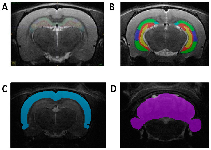
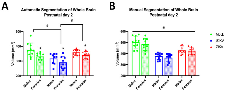
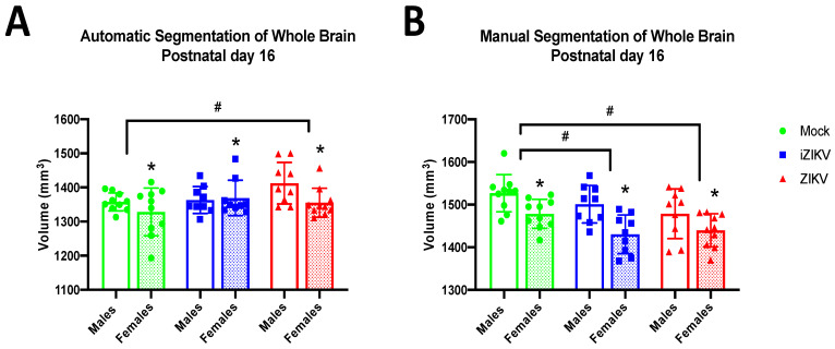
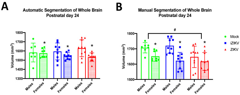
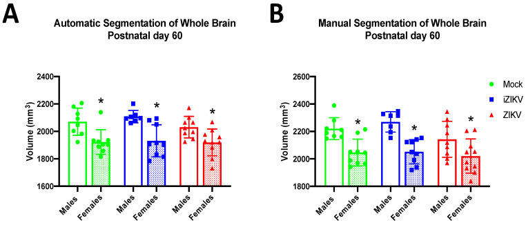
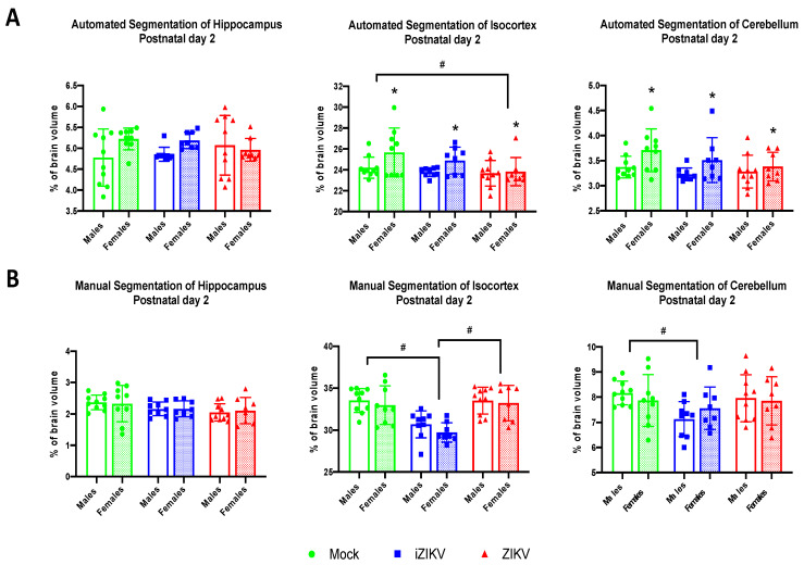
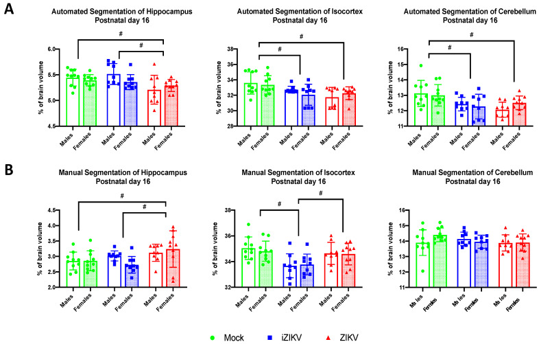
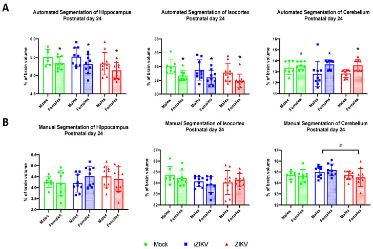
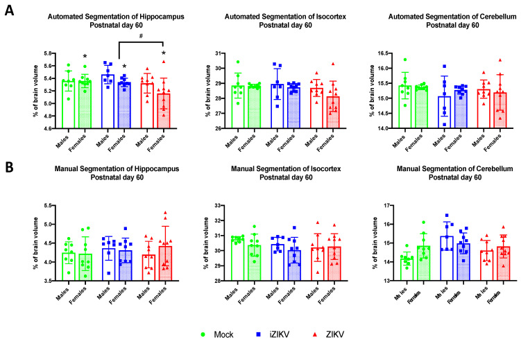
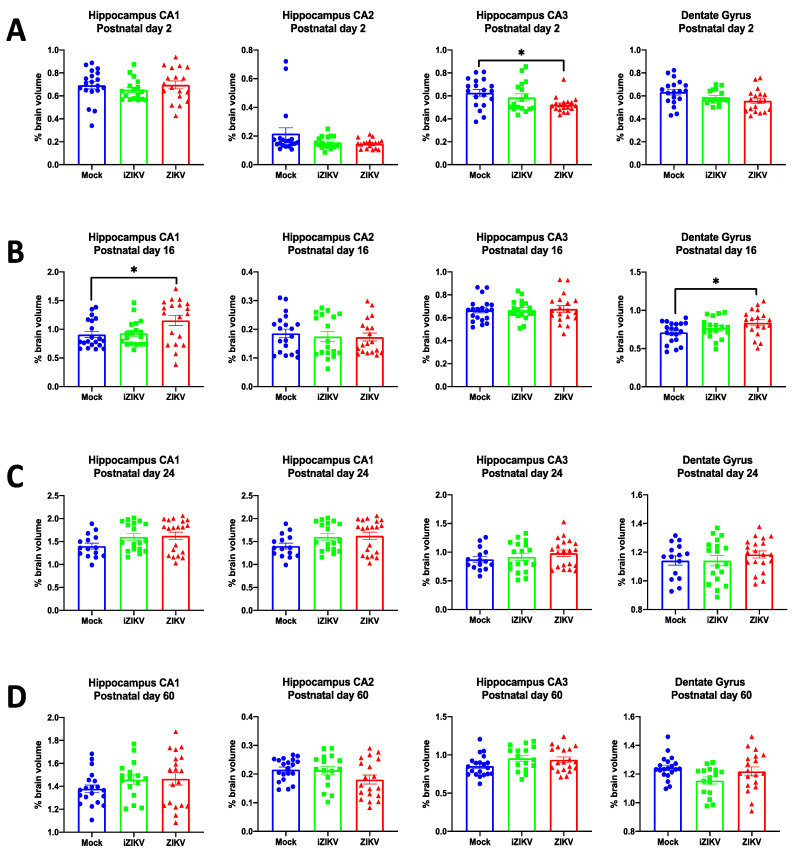
Since Zika virus (ZIKV) first emerged as a public health concern in 2015, our ability to identify and track the long-term neurological sequelae of prenatal Zika virus (ZIKV) infection in humans has been limited. Our lab has developed a rat model of maternal ZIKV infection with associated vertical transmission to the fetus that results in significant brain malformations in the neonatal offspring. Here, we use this model in conjunction with longitudinal magnetic resonance imaging (MRI) to expand our understanding of the long-term neurological consequences of prenatal ZIKV infection in order to identify characteristic neurodevelopmental changes and track them across time. We exploited both manual and automated atlas-based segmentation of MR images in order to identify long-term structural changes within the developing rat brain following inoculation. The paradigm involved scanning three cohorts of male and female rats that were prenatally inoculated with 107 PFU ZIKV, 107 UV-inactivated ZIKV (iZIKV), or diluent medium (mock), at 4 different postnatal day (P) age points: P2, P16, P24, and P60. Analysis of tracked brain structures revealed significantly altered development in both the ZIKV and iZIKV rats. Moreover, we demonstrate that prenatal ZIKV infection alters the growth of brain regions throughout the neonatal and juvenile ages. Our findings also suggest that maternal immune activation caused by inactive viral proteins may play a role in altered brain growth throughout development. For the very first time, we introduce manual and automated atlas-based segmentation of neonatal and juvenile rat brains longitudinally. Experimental results demonstrate the effectiveness of our novel approach for detecting significant changes in neurodevelopment in models of early-life infections.

Keywords: MRI; Zika virus (ZIKV); congenital infection; neurodevelopment; neuroimaging; pregnancy.


Conflict of interest statement

P.K. has financial interests in Ekam Solutions, the company that developed the Ekam Visualization and Analysis (EVA) tool used in this study. The other authors declare no conflicts of interest.

Figures

Figure 1
Figure 1
Representative images of manual segmentation in the coronal plane at P60. (A) Delineation of hippocampus and its subregions segmented at a more rostral level. (B) Delineation of hippocampus and its subregions segmented at a more caudal level. (C) Depiction of the isocortex region manually segmented. (D) Depiction of the cerebellum region manually segmented.
Figure 2
Figure 2
Absolute whole-brain volumes in postnatal day 2 neonates across all three groups stratified by sex using both automated and manual methods of segmentation (n = 8–10 per sex per group). (A) Automated segmentation revealed a main effect of inoculation (# p  <  0.05). Specifically, whole-brain volume was significantly smaller in iZIKV pups when compared to both mock control and ZIKV pups (post-hoc, # p  <  0.05). Analysis also revealed a main effect of sex (* p  <  0.05); P2 females across all groups had significantly smaller whole-brain volumes than their male counterparts. No interaction between inoculation and sex was detected. (B) Manual segmentation also revealed a main effect of inoculation (# p  <  0.05). iZIKV pups again displayed significantly reduced whole-brain volumes when compared with all other groups; however, ZIKV pups also displayed significantly reduced whole-brain volumes when compared to mock controls (post-hoc, # p  <  0.05). There was no main effect of sex or interaction of inoculation × sex. Bars represent the mean ± SD and data points represent individual subjects. Data were analyzed using a two-way ANOVA with inoculation (# p  <  0.05) and sex (* p  <  0.05) as factors, followed by Tukey’s test.
Figure 3
Figure 3
Absolute whole-brain volumes in postnatal day 16 juveniles across all three groups stratified by sex using both automated and manual methods of segmentation (n = 9–10 per sex per group). (A) Automated segmentation revealed a main effect of inoculation (# p < 0.05). Specifically, whole-brain volume was significantly larger in ZIKV juveniles when compared to the mock control group (post-hoc, # p < 0.05). (B) Manual segmentation also revealed a main effect of inoculation (# p < 0.05). Both iZIKV and ZIKV juveniles displayed significantly reduced whole-brain volumes when compared to mock control juveniles (post-hoc, # p < 0.05). Analyses also revealed a main effect of sex for both segmentation methods (* p < 0.05). More specifically, P16 females across all groups had significantly smaller whole-brain volumes than their male counterparts. There was no interaction of inoculation × sex within either method of segmentation. Bars represent the mean ± SD and data points represent individual subjects. Data were analyzed using a two-way ANOVA with inoculation (# p < 0.05) and sex (* p < 0.05) as factors, followed by Tukey’s test.
Figure 4
Figure 4
Absolute whole-brain volumes in postnatal day 24 juveniles across all three groups stratified by sex using both methods of segmentation (n = 7–10 per sex per group). (A) Automated segmentation revealed no main effect of inoculation. (B) Manual segmentation revealed a main effect of inoculation (# p  <  0.05). ZIKV juveniles displayed significantly reduced whole-brain volumes when compared to mock control juveniles (post-hoc, # p  <  0.05). Analyses also revealed a main effect of sex for both segmentation methods (* p  <  0.05). More specifically, P24 females across all groups had significantly smaller whole-brain volumes than their male counterparts. There was no interaction of inoculation × sex within either method of segmentation. Bars represent the mean  ±  SD and data points represent individual subjects. Data were analyzed using a two-way ANOVA with inoculation (# p  <  0.05) and sex (* p  <  0.05) as factors, followed by Tukey’s test.
Figure 5
Figure 5
Absolute whole-brain volumes in postnatal day 60 adults across all three groups stratified by sex using both automated segmentation (A) and manual segmentation (B), (n = 7–10 per sex per group). Neither method detected a main effect of inoculation. Within both segmentation methods, analyses revealed a main effect of sex (* p  <  0.05). More specifically, P60 females across all groups had significantly smaller whole-brain volumes than their male counterparts. There was no interaction of inoculation × sex within either method of segmentation. Bars represent the mean  ±  SD and data points represent individual subjects. Data were analyzed using a two-way ANOVA with inoculation and sex (* p < 0.05) as factors, followed by Tukey’s test.
Figure 6
Figure 6
(A) Automated segmentation of brain regions at postnatal day 2 revealed a significant effect of inoculation. Analysis revealed ZIKV-inoculated neonates had a smaller isocortex when compared to mock-inoculated neonates (post-hoc,# p < 0.05). Furthermore, analysis revealed a significant effect of sex in both the isocortex and cerebellum (* p < 0.05). Analysis detected no interaction of inoculation × sex. (B) Manual segmentation of brain regions at postnatal day 2 revealed a significant effect of inoculation. Within the isocortex, iZIKV-inoculated neonates displayed smaller regional volumes when compared to both mock and ZIKV counterparts (post-hoc,# p < 0.05). Analysis also revealed iZIKV neonates had smaller cerebellums in comparison to the mock controls (post-hoc,# p < 0.05). Analysis detected no significant effect of sex or interaction of inoculation × sex. Bars represent the mean ± SD and data points represent individual subjects (n = 8–10 per sex per group). Data were analyzed using a two-way ANOVA with inoculation (# p < 0.05) and sex (* p < 0.05) as factors, followed by Tukey’s test.
Figure 7
Figure 7
(A) Automated segmentation of brain regions at postnatal day 16 revealed a significant effect of inoculation. Within the hippocampus region, ZIKV-inoculated juveniles displayed smaller regional volumes when compared to both mock and iZIKV counterparts (post-hoc,# p < 0.05). Within the isocortex and cerebellum, both iZIKV-inoculated and ZIKV-inoculated juveniles displayed smaller regional volumes when compared to mock controls (post-hoc,# p < 0.05). Analysis detected no significant effect of sex or interaction of inoculation × sex. (B) Manual segmentation of brain regions at postnatal day 16 revealed a significant effect of inoculation. Within the hippocampus region, ZIKV-inoculated juveniles displayed larger regional volumes when compared to both mock and iZIKV counterparts (post-hoc,# p < 0.05). Within the isocortex, iZIKV-inoculated juveniles displayed smaller regional volumes when compared to both mock and ZIKV counterparts (post-hoc, # p < 0.05). Analysis detected no significant effect of sex or interaction of inoculation × sex. Bars represent the mean ± SD and data points represent individual subjects (n = 9–10 per sex per group). Data were analyzed using a two-way ANOVA with inoculation (# p < 0.05) and sex as factors, followed by Tukey’s test.
Figure 8
Figure 8
(A) Automated segmentation of brain regions for postnatal day 24 revealed a main effect of sex in all regions analyzed (* p < 0.05). Furthermore, analysis identified no significant effect of inoculation or interaction of inoculation × sex. (B) Manual segmentation of brain regions at postnatal day 24 revealed a significant effect of inoculation. Within the cerebellum region, ZIKV-inoculated juveniles displayed smaller regional volumes when compared to the iZIKV-inoculated juveniles (post-hoc,# p < 0.05). Analysis detected no significant effect of sex or interaction of inoculation × sex. Bars represent the mean ± SD and data points represent individual subjects (n = 7–10 per sex per group). Data were analyzed using a two-way ANOVA with inoculation (# p < 0.05) and sex (* p < 0.05) as factors, followed by Tukey’s test.
Figure 9
Figure 9
(A) Automated segmentation of brain regions at postnatal day 60 revealed a significant effect of inoculation. Within the hippocampus region, ZIKV-inoculated juveniles displayed smaller regional volumes when compared to the iZIKV-inoculated juveniles (post-hoc,# p < 0.05). Moreover, analysis also revealed a main effect of sex within the hippocampal region (* p < 0.05). Analysis detected no interaction of inoculation × sex. (B) Analysis of manually segmented brain regions for postnatal day 60 revealed no significant effect of inoculation, sex, or interaction of inoculation × sex. Bars represent the mean ± SD and data points represent individual subjects (n = 7–10 per sex per group). Data were analyzed using a two-way ANOVA with inoculation (# p < 0.05) and sex (* p < 0.05) as factors, followed by Tukey’s test.
Figure 10
Figure 10
Analysis of manually segmented hippocampal subregions for postnatal day 2 (n = 17–19 per group) (A), postnatal day 16 (n = 18–20 per group) (B), postnatal day 24 (n = 15–20 per group) (C), and postnatal day 60 (n = 16–19 per group) (D). At P2, volumetric analysis of hippocampal subregions revealed a significant effect of inoculation within the CA3 of the hippocampus. Specifically, the ZIKV-inoculated pups displayed a significant volume reduction in the CA3 subregion when compared to mock counterparts (post-hoc, * p < 0.05). At P16, analysis revealed a significant effect of inoculation within the CA1 of the hippocampus and dentate gyrus. Specifically, ZIKV-inoculated juveniles displayed significantly larger volumes in both the CA1 subregion and dentate gyrus when compared to mock controls (post-hoc, * p < 0.05). Across all groups, bars represent the mean ± SD in each subregion of the hippocampus while data points represent each individual rat.

References

    1. Valentine M.J., Murdock C.C., Kelly P.J. Sylvatic Cycles of Arboviruses in Non-Human Primates. Parasit. Vectors. 2019;12:463. doi: 10.1186/s13071-019-3732-0. - DOI - PMC - PubMed
    1. Braack L., Gouveia de Almeida A.P., Cornel A.J., Swanepoel R., de Jager C. Mosquito-Borne Arboviruses of African Origin: Review of Key Viruses and Vectors. Parasit. Vectors. 2018;11 doi: 10.1186/s13071-017-2559-9. - DOI - PMC - PubMed
    1. Gregory C.J., Oduyebo T., Brault A.C., Brooks J.T., Chung K.-W., Hills S., Kuehnert M.J., Mead P., Meaney-Delman D., Rabe I., et al. Modes of Transmission of Zika Virus. J. Infect. Dis. 2017;216:S875–S883. doi: 10.1093/infdis/jix396. - DOI - PMC - PubMed
    1. Mann T.Z., Haddad L.B., Williams T.R., Hills S.L., Read J.S., Dee D.L., Dziuban E.J., Pérez-Padilla J., Jamieson D.J., Honein M.A., et al. Breast Milk Transmission of Flaviviruses in the Context of Zika Virus: A Systematic Review. Paediatr. Perinat. Epidemiol. 2018;32:358–368. doi: 10.1111/ppe.12478. - DOI - PMC - PubMed
    1. Sharma A., Lal S.K. Zika Virus: Transmission, Detection, Control, and Prevention. Front. Microbiol. 2017;8 doi: 10.3389/fmicb.2017.00110. - DOI - PMC - PubMed

Publication types

MeSH terms