. 2021 Jun 11;11(6):1553.
doi: 10.3390/nano11061553.

Two Approaches to the Laser-Induced Formation of Au/Ag Bimetallic Nanoparticles in Supercritical Carbon Dioxide

Affiliations

Two Approaches to the Laser-Induced Formation of Au/Ag Bimetallic Nanoparticles in Supercritical Carbon Dioxide

Alexey Rybaltovsky et al. Nanomaterials (Basel). .

Abstract

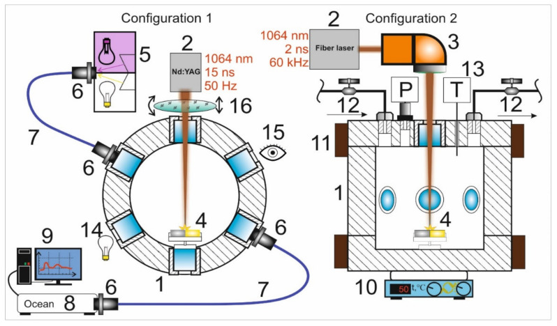
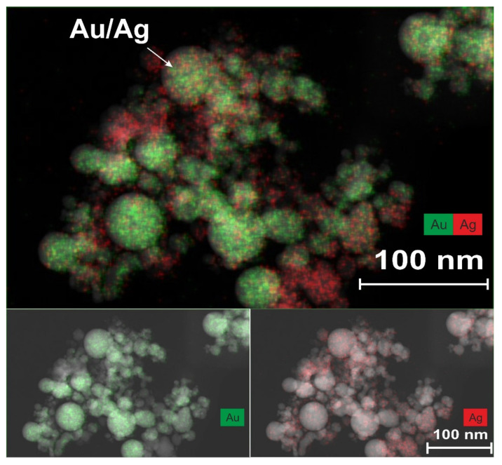
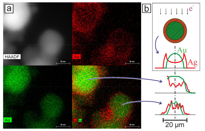
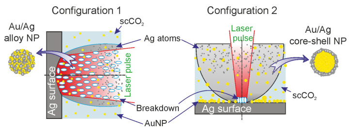
Two approaches are proposed for the synthesis of bimetallic Au/Ag nanoparticles, using the pulsed laser ablation of a target consisting of gold and silver plates in a medium of supercritical carbon dioxide. The differences between the two approaches related to the field of "green chemistry" are in the use of different geometric configurations and different laser sources when carrying out the experiments. In the first configuration, the Ag and Au targets are placed side-by-side vertically on the side wall of a high-pressure reactor and the ablation of the target plates occurs alternately with a stationary "wide" horizontal beam with a laser pulse repetition rate of 50 Hz. In the second configuration, the targets are placed horizontally at the bottom of a reactor and the ablation of their parts is carried out by scanning from above with a vertical "narrow" laser beam with a pulse repetition rate of 60 kHz. The possibility of obtaining Ag/Au alloy nanoparticles is demonstrated using the first configuration, while the possibility of obtaining "core-shell" bimetallic Au/Ag nanoparticles with a gold core and a silver shell is demonstrated using the second configuration. A simple model is proposed to explain the obtained results.

Keywords: laser ablation; plasmonic nanoparticles; supercritical carbon dioxide; supercritical fluid.


Conflict of interest statement

The authors declare no conflict of interest.

Figures

Figure 1
Figure 1
Schemes of installations for the synthesis of Ag/Au BMNPs show two different models of ablation. The schematic sections of the reactor, showing top (for configuration 1) and side views (for configuration 2): 1—high-pressure reactor; 2—laser sources; 3—galvo scanner with F-theta lens; 4—gold and silver targets; 5—UV and visible light sources; 6—collimator lens; 7—optical fibers; 8—spectrometer; 9—PC; 10—heating plate; 11—ring heaters; 12—needle valves for CO2 inlet and outlet; 13—pressure and temperature sensors; 14—backlight; 15—observation window; 16—quartz lens.
Figure 2
Figure 2
(a) Transformation of the extinction spectra of a colloidal solution of scCO2 during successive cycles of ablation of Au and Ag plates in configuration 1. The numbers corresponding to the curves show the times since the beginning of the ablation of the indicated (Au or Ag) target. (b) Variant of decomposition of the final spectrum (red line) into the sum (black dashed line) of individual Gaussian components (1–4) and the Rayleigh scattering component (Rayleigh).
Figure 3
Figure 3
Transformation of the extinction spectra of a colloidal solution scCO2 during successive cycles of ablation of the (a) gold and (b) the silver parts of the target. (c) Simultaneous ablation of both parts in configuration 2. The numbers on the curves show the times from the beginning of the ablation in seconds. The numbers to the right of the curves show the times in seconds since the start of the ablation cycle. (d) Variation of the decomposition of the spectrum obtained 10 s after the end of the last stage of ablation (red line) into the sum (black dashed line) of individual Gaussian components (1–4) and the Rayleigh scattering component (Rayleigh).
Figure 4
Figure 4
TEM images of nanoparticles obtained using the EDX method. The arrow indicates one of the largest Ag/Au-BMNP-type “alloys”, measuring up to 40 nm in size. Nanoparticles were synthesized in the setup of configuration 1, using a low-frequency laser source with a high pulse energy.
Figure 5
Figure 5
(a) TEM images of Au/Ag “core–shell” BMNPs, demonstrated using the EDX technique. Nanoparticles were obtained by laser synthesis in configuration N2. (b) Top: A cross-sectional image of a nanoparticle model with a core–shell structure and the pixel intensity profile of the elements from the core and shell for the corresponding EDX TEM image. Arrows show the direction of the flow of electrons (e-) during image acquisition. Bottom: Pixel intensity profiles for Au and Ag for TEM images of two nanoparticles.
Figure 6
Figure 6
Model representation of the processes occurring during the ablation of a silver target in two different configuration setups (Figure 1) and leading to the synthesis of Au/Ag BMNP “alloy” (configuration 1) and “core–shell” (configuration 2) types.

References

    1. Jellinek J. Nanoalloys: Tuning properties and characteristics through size and composition. Faraday Discuss. 2008;138:11–35. doi: 10.1039/b800086g. - DOI - PubMed
    1. Eng N.B. Synergetic Antibacterial Effects of Silver Nanoparticles @ Aloe Vera Prepared via a Green Method. Nano Biomed. Eng. 2010;2:267–274. doi: 10.5101/nbe.v2i4.p252-257.1. - DOI
    1. Meena Kumari M., Jacob J., Philip D. Green synthesis and applications of Au-Ag bimetallic nanoparticles. Spectrochim. Acta Part A Mol. Biomol. Spectrosc. 2015;137:185–192. doi: 10.1016/j.saa.2014.08.079. - DOI - PubMed
    1. Boote B.W., Byun H., Kim J.H. Silver-gold bimetallic nanoparticles and their applications as optical materials. J. Nanosci. Nanotechnol. 2014;14:1563–1577. doi: 10.1166/jnn.2014.9077. - DOI - PubMed
    1. Intartaglia R., Das G., Bagga K., Gopalakrishnan A., Genovese A., Povia M., di Fabrizio E., Cingolani R., Diaspro A., Brandi F. Laser synthesis of ligand-free bimetallic nanoparticles for plasmonic applications. Phys. Chem. Chem. Phys. 2013;15:3075–3082. doi: 10.1039/C2CP42656K. - DOI - PubMed

LinkOut - more resources