. 2021 Jun 11;11(6):1077.
doi: 10.3390/diagnostics11061077.

Deep Learning-Based Post-Processing of Real-Time MRI to Assess and Quantify Dynamic Wrist Movement in Health and Disease

Affiliations

Deep Learning-Based Post-Processing of Real-Time MRI to Assess and Quantify Dynamic Wrist Movement in Health and Disease

Karl Ludger Radke et al. Diagnostics (Basel). .

Abstract

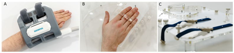
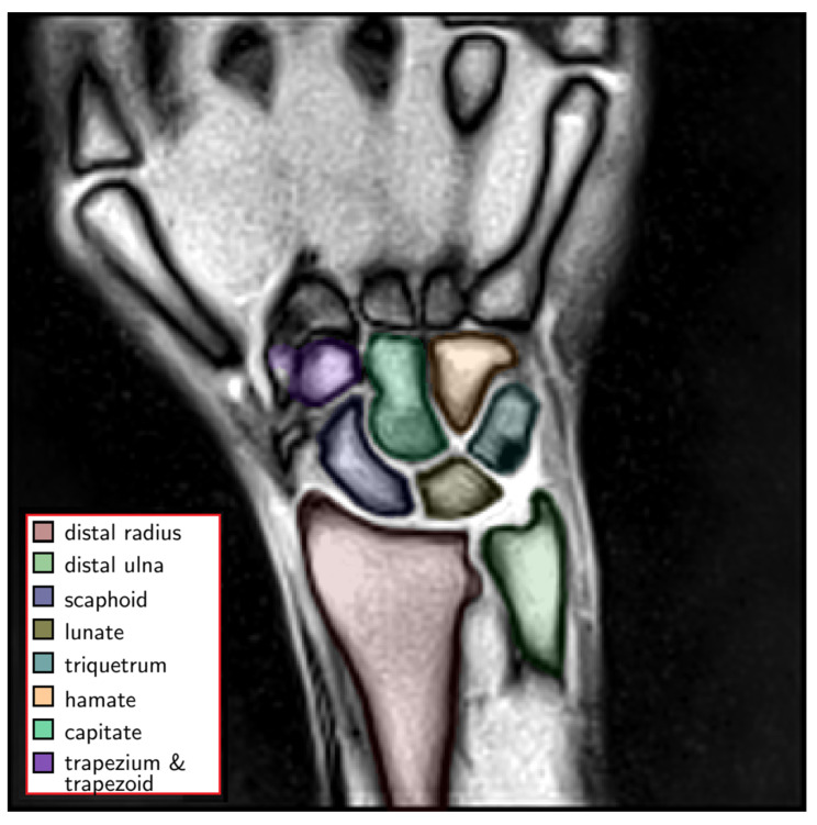
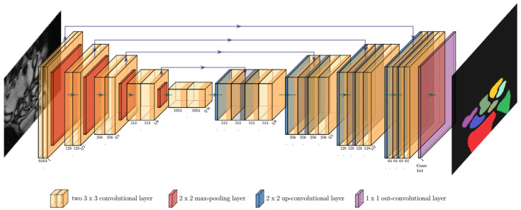
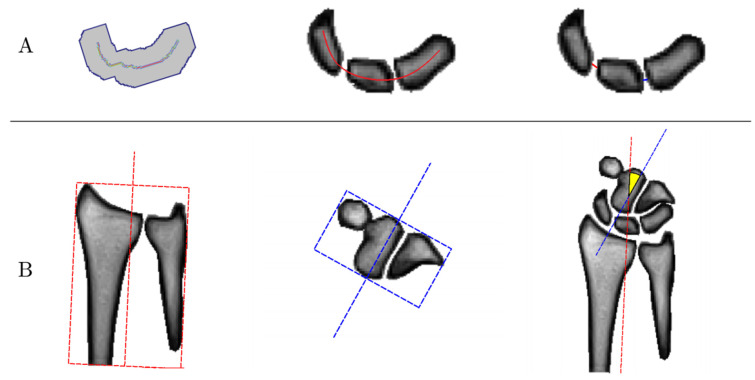
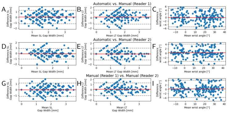
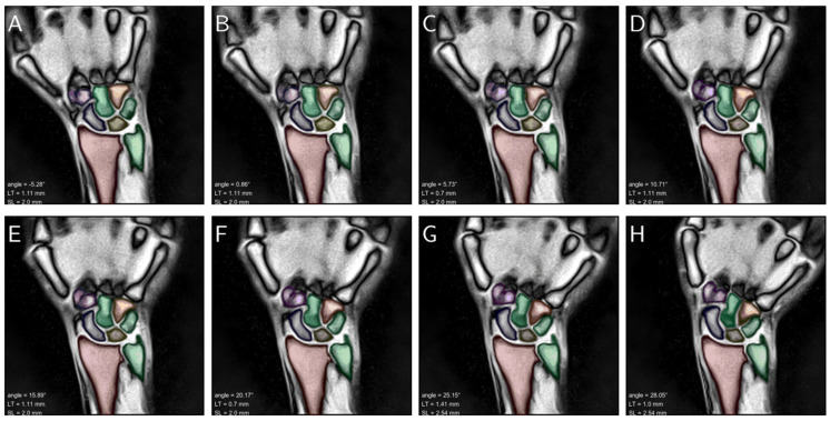
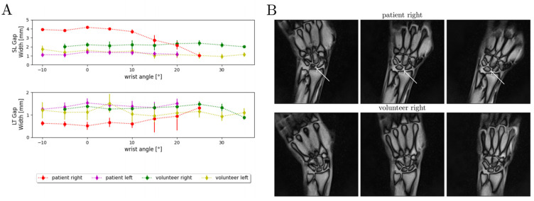
While morphologic magnetic resonance imaging (MRI) is the imaging modality of choice for the evaluation of ligamentous wrist injuries, it is merely static and incapable of diagnosing dynamic wrist instability. Based on real-time MRI and algorithm-based image post-processing in terms of convolutional neural networks (CNNs), this study aims to develop and validate an automatic technique to quantify wrist movement. A total of 56 bilateral wrists (28 healthy volunteers) were imaged during continuous and alternating maximum ulnar and radial abduction. Following CNN-based automatic segmentations of carpal bone contours, scapholunate and lunotriquetral gap widths were quantified based on dedicated algorithms and as a function of wrist position. Automatic segmentations were in excellent agreement with manual reference segmentations performed by two radiologists as indicated by Dice similarity coefficients of 0.96 ± 0.02 and consistent and unskewed Bland-Altman plots. Clinical applicability of the framework was assessed in a patient with diagnosed scapholunate ligament injury. Considerable increases in scapholunate gap widths across the range-of-motion were found. In conclusion, the combination of real-time wrist MRI and the present framework provides a powerful diagnostic tool for dynamic assessment of wrist function and, if confirmed in clinical trials, dynamic carpal instability that may elude static assessment using clinical-standard imaging modalities.

Keywords: carpal instability; deep learning; dynamic instability; magnetic resonance imaging; real-time; scapholunate ligament injury.


Conflict of interest statement

The authors declare no conflict of interest. The funders had no role in the design of the study; in the collection, analyzes, or interpretation of data; in the writing of the manuscript, or in the decision to publish the results.

Figures

Figure 1
Figure 1
Coil and measurement setups during static reference and dynamic real-time MRI measurements. (A) For static measurements, a four-channel flex coil (Siemens Healthineers) was wrapped around the wrist to acquire the morphologic sequences. Shown are a left forearm and hand that were immobilized and imaged for reference purposes. (B) Positioning of a right wrist on the custom-made MRI-compatible movement device. The device has a mobile sliding tray (§) with a predefined range and trajectory of movement (x) onto which the hand was placed. (C) Tourniquets (*) were used to fix the forearm and prevent adaptive movement. The underside of the mobile sliding tray was coated with synthetic polytetrafluoroethylene (“Teflon”) to reduce friction. Centered underneath the wrist is the pivot point (#), which connects the mobile sliding tray and the fixed base plate. For dynamic measurements, a 24-channel spine matrix coil and an 18-channel body coil (both from Siemens Healthineers) were positioned underneath (spine matrix coil) and on top of the device (body coil) [not shown].
Figure 2
Figure 2
Exemplary color-coded manual segmentations of the wrist bones, i.e., carpal bones and distal forearm (left). These manual segmentations served as the ground truth for subsequent automatization. Uncolored anatomic structures are not part of the wrist and were considered as background.
Figure 3
Figure 3
Convolution of an input MR image (left) inside the U-Net architecture and visualization of resultant bone segmentations as its output (right). The number of channels is indicated under each convolutional layer. The reduction and increase of the image resolution are indicated at the end of a block normalized to I = input size. Color-codes of the output image correspond to the different classes of semantic segmentations.
Figure 4
Figure 4
Graphical visualization of the algorithm-based measurements of carpal configuration. (A) Determination of scapholunate (SL) and lunotriquetral (LT) gap widths. First, the bone masks of the scaphoid, lunate, and triquetrum were connected (left). Then, the centerline of the three bones was determined along the carpal arcs (center). Eventually, the SL (red) and LT (blue) gap widths were determined based on the intersections of this centerline and the bone contours. (B) Determination of the wrist angle. The minimum bounding boxes around the bone contours of the forearm, i.e., radius and ulna (red, left), as well as around the distal row of the carpal bones (blue, center) were determined. Then, the wrist angle was calculated as the angle between the centers of the two boxes (yellow, right).
Figure 5
Figure 5
Bland–Altman plots to evaluate inter-method and inter-reader reliability. Automatic segmentations and subsequently determined diagnostic measures, i.e., scapholunate (SL) gap width (A,D,G), lunotriquetral (LT) gap width (B,E,H), and wrist angle (C,F,I), were evaluated against the manual reference measurements by two radiologists (reader 1, reader 2). Y-axes indicate the respective measures’ inter-method or inter-reader differences, while x-axes indicate the mean measure. Visualized are the 210 values of the test datasets as derived from 15 representative images across the entire range-of-motion. Red lines indicate the mean of the differences and gray dashed lines the 95% confidence intervals.
Figure 6
Figure 6
Consecutive still images of a left wrist across the entire range of active radioulnar movement that ranged from maximum radial abduction to maximum ulnar abduction (AH). The carpus and forearm are color-coded as in Figure 2 and overlaid onto the morphologic images. Inset (lower left) are the resultant wrist angles (“angle”), LT gap width (“LT”), and SL gap width (“SL”). The corresponding video that visualizes fluent movement across the entire range-of-motion is appended as Video S1.
Figure 7
Figure 7
Quantitative and qualitative changes in carpal configurations in a patient with a partial scapholunate ligament rupture and a gender- and size-matched healthy volunteer during active radioulnar movement. (A) SL and LT gap widths are given as a function of wrist angle across the range-of-motion. The gap widths are indicated as means (dots) and standard deviations (whiskers). A total of 300 MR images per wrist were analyzed and quantified by our framework. Indicated are the measured SL and LT gap widths of the wrist-injured patient (partial SL ligament rupture, “patient right”, red), the contralateral wrist (“patient left”, purple), and the corresponding wrists of the matched and healthy volunteer (“volunteer right”, green; “volunteer left”, yellow). For the sake of visualization, the wrist angles are grouped at intervals of 5°. (B) Three exemplary MR images are shown for the patient and the volunteer at various positions throughout the active radioulnar movement range. The increased dehiscence of the SL gap of the injured wrist (white arrows) is particularly obvious in the neutral position and ulnar abduction and at the proximal portion of the SL gap.

References

    1. Shaw C.B., Foster B.H., Borgese M., Boutin R.D., Bateni C., Boonsri P., Bayne C.O., Szabo R.M., Nayak K.S., Chaudhari A.J. Real-time three-dimensional MRI for the assessment of dynamic carpal instability. PLoS ONE. 2019;14:e0222704. doi: 10.1371/journal.pone.0222704. - DOI - PMC - PubMed
    1. Schernberg F. Roentgenographic examination of the wrist: A systematic study of the normal, lax and injured wrist Part 2: Stress views. J. Hand Surg. 1990;15:220–228. doi: 10.1016/0266-7681(90)90127-P. - DOI - PubMed
    1. Moser T., Dosch J.-C., Moussaoui A., Dietemann J.-L. Wrist Ligament Tears: Evaluation of MRI and Combined MDCT and MR Arthrography. Am. J. Roentgenol. 2007;188:1278–1286. doi: 10.2214/AJR.06.0288. - DOI - PubMed
    1. Theumann N.H., Etechami G., Duvoisin B., Wintermark M., Schnyder P., Favarger N., Gilula L.A. Association between Extrinsic and Intrinsic Carpal Ligament Injuries at MR Arthrography and Carpal Instability at Radiography: Initial Observations. Radiology. 2006;238:950–957. doi: 10.1148/radiol.2383050013. - DOI - PubMed
    1. Watson H.K., Ballet F.L. The SLAC wrist: Scapholunate advanced collapse pattern of degenerative arthritis. J. Hand Surg. 1984;9:358–365. doi: 10.1016/S0363-5023(84)80223-3. - DOI - PubMed

LinkOut - more resources