Review
. 2021 Jun 16;13(12):3021.
doi: 10.3390/cancers13123021.

Pitfalls in the Diagnosis of Nodular Lymphocyte Predominant Hodgkin Lymphoma: Variant Patterns, Borderlines and Mimics

Affiliations
Review

Pitfalls in the Diagnosis of Nodular Lymphocyte Predominant Hodgkin Lymphoma: Variant Patterns, Borderlines and Mimics

Sheren Younes et al. Cancers (Basel). .

Abstract

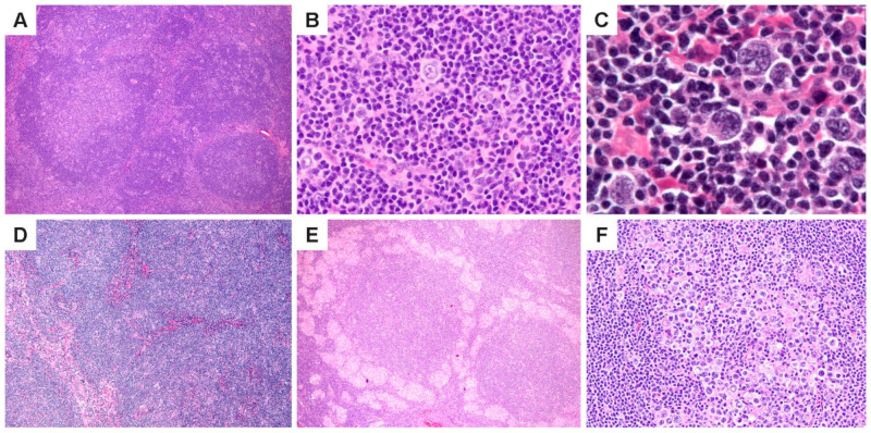
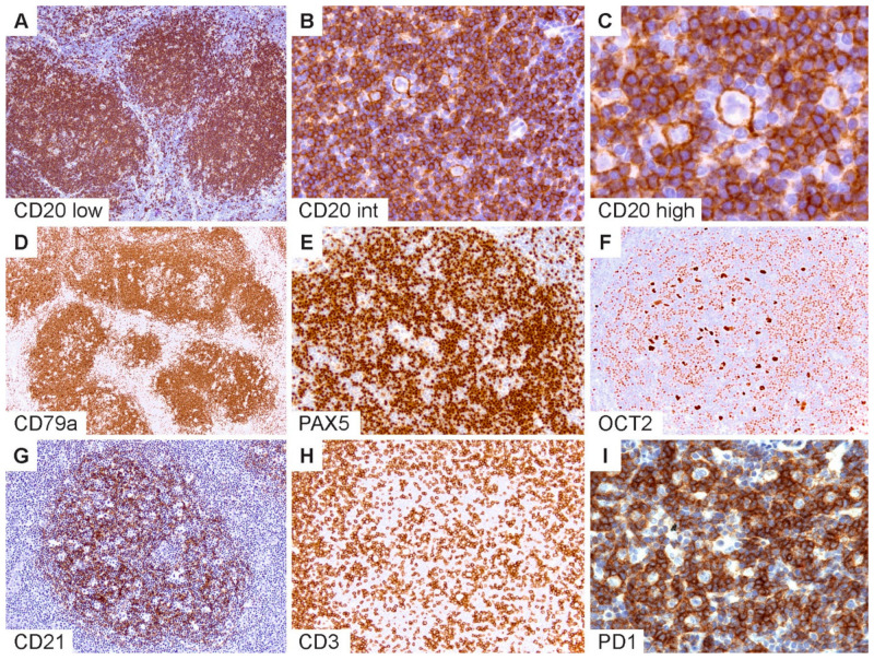
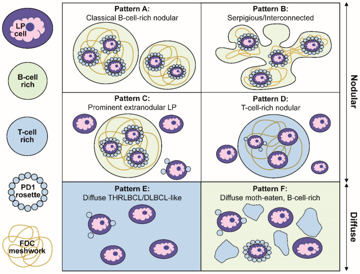
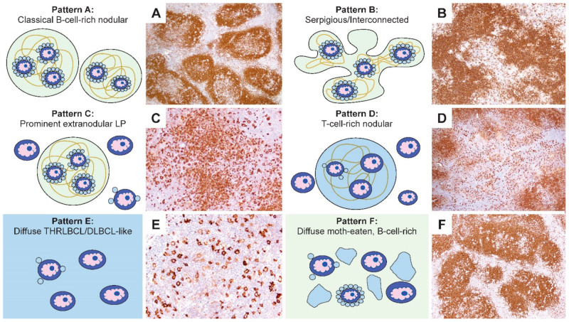
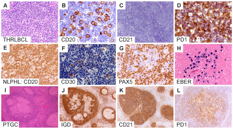
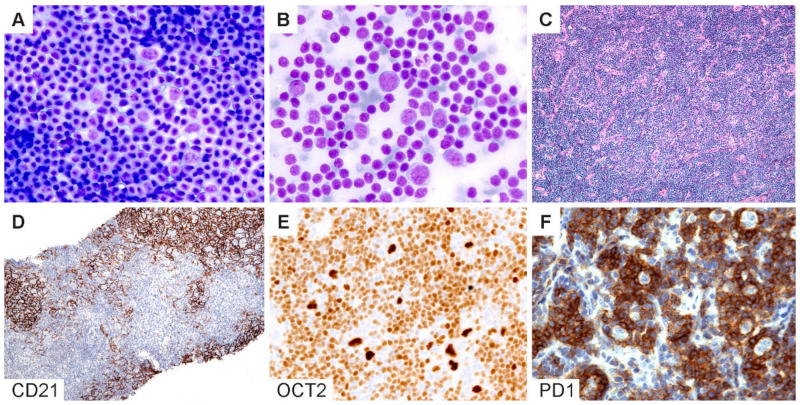
Nodular lymphocyte predominant Hodgkin lymphoma (NLPHL) represents approximately 5% of Hodgkin lymphoma and typically affects children and young adults. Although the overall prognosis is favorable, variant growth patterns in NLPHL correlate with disease recurrence and progression to T-cell/histiocyte-rich large B-cell lymphoma or frank diffuse large B-cell lymphoma (DLBCL). The diagnostic boundary between NLPHL and DLBCL can be difficult to discern, especially in the presence of variant histologies. Both diagnoses are established using morphology and immunophenotype and share similarities, including the infrequent large tumor B-cells and the lymphocyte and histiocyte-rich microenvironment. NLPHL also shows overlap with other lymphomas, particularly, classic Hodgkin lymphoma and T-cell lymphomas. Similarly, there is overlap with non-neoplastic conditions, such as the progressive transformation of germinal centers. Given the significant clinical differences among these entities, it is imperative that NLPHL and its variants are carefully separated from other lymphomas and their mimics. In this article, the characteristic features of NLPHL and its diagnostic boundaries and pitfalls are discussed. The current understanding of genetic features and immune microenvironment will be addressed, such that a framework to better understand biological behavior and customize patient care is provided.

Keywords: Hodgkin lymphoma; LP cell; T-cell/histocyte-rich large B-cell lymphoma; diffuse large B-cell lymphoma; lymphocyte predominant; microenvironment.


Conflict of interest statement

The authors declare no conflict of interest.

Figures

Figure 1
Figure 1
Histologic features of NLPHL. (A) Lymph node effaced by a proliferation of classic lymphoid nodules. (B,C), Scattered LP cells in a microenvironment rich in small lymphocytes and histiocytes. (D) Nodules interspersed by bands of fibrosis. (E) Nodules surrounded by epithelioid histiocytes. (F) A nodule with increased LP cells. [Original magnifications: A,D,E ×60; B,F ×150; C ×600].
Figure 2
Figure 2
Immunophenotypic features of NLPHL. (A) CD20 highlights numerous classic lymphoid nodules. (B,C) Scattered LP cells reside within nodules containing a microenvironment rich in small B-lymphocytes. (D) CD79a, (E) PAX5 and (F) OCT2 show staining of LP cells and the surrounding small B cells. (G) CD21 defines an intact follicular dendritic cell meshwork within an NLPHL nodule. (H) CD3 stains background T cells with occasional ring formations and (I) prominent PD1-positive rings surrounding LP cells. [Original magnifications: A,D,G,H ×60; B,E,F,I ×150; C ×600].
Figure 3
Figure 3
Schematic representation of the six immunoarchitectural patterns of NLPHL. (A) Classical B-cell-rich noduless and (B) serpiginous/interconnected nodules define the typical patterns of NLPHL, with most LP cells residing within the nodules associated with FDC meshworks. (C) Prominent extranodular LP cells define pattern C, whereas (D) T-cell-rich nodules define pattern D. (E) The diffuse THRLBCL/DLBCL-like pattern and (F) diffuse moth-eaten B-cell-rich pattern show diffuse growths unassociated with the FDC meshworks.
Figure 4
Figure 4
Comparison of the schematic representations and CD20-stained NLPHL patterns. (AF) Representative CD20-stained examples of the six NLPHL patterns highlight the number and localization of the LP cells, as well as the content and configuration of small B cells and T cells in the microenvironment in patterns AF. [Original magnifications: A,B,D,F ×60; C,E ×150].
Figure 5
Figure 5
Diagnostic boundaries surrounding NLPHL. (AD) T/histiocyte-rich large B-cell lymphomas show diffuse growth, scattered LP cells and an absence of FDC meshworks. (EH) NLPHL mimic of lymphocyte-rich classic Hodgkin lymphoma shows the expression of CD20, CD30, dim PAX5 and EBV within LP cells. (IL) Progressive transformation of germinal centers show involution of the IgD-positive mantle zone B-cells associated with CD21-positive FDC meshworks and an atypical distribution of PD1-positive T cells in an affected nodule. [Original magnifications: A,C,I,J,K ×60; B,D,E,F,G,H,L ×150].
Figure 6
Figure 6
Cytologic diagnosis of NLPHL. (A,B) Wright–Giemsa-stained touch preparations show LP cells in a background of small lymphoid cells and occasional inflammatory cells. (C) H&E section of a core needle biopsy does not readily reveal a nodular architecture. (D) A CD21 stain highlights the FDC meshworks and confirms the presence of nodules. (E) Bright OCT2 staining and (F) prominent PD1-positive rings lend support for the diagnosis of NLPHL. [Original magnifications: A,B ×600; C,D ×40; E,F ×150].

References

    1. Hansmann M.-L., Zwingers T., Lennert K. Clinical features of nodular paragranuloma (Hodgkin’s disease, lymphocyte predominance type, nodular) J. Cancer Res. Clin. Oncol. 1984;108:321–330. doi: 10.1007/BF00390466. - DOI - PMC - PubMed
    1. Morton L.M., Wang S.S., Devesa S.S., Hartge P., Weisenburger D.D., Linet M.S. Lymphoma incidence patterns by WHO subtype in the United States, 1992–2001. Blood. 2006;107:265–276. doi: 10.1182/blood-2005-06-2508. - DOI - PMC - PubMed
    1. Teras L.R., DeSantis C.E., Cerhan J.R., Morton L.M., Jemal A., Flowers C.R. 2016 US lymphoid malignancy statistics by World Health Organization subtypes. CA Cancer J. Clin. 2016;66:443–459. doi: 10.3322/caac.21357. - DOI - PubMed
    1. Stein H., Swerdlow S.H. Nodular lymphocyte predominant Hodgkin lymphoma. In: Swerdlow S.H., Campo E., Harris N.L., Jaffe E.S., Pileri S.A., Stein H., Thiele J., editors. WHO Classification of Tumours of Haematopoietic and Lymphoid Tissues. IARC Press; Lyon, France: 2017.
    1. Appel B.E., Chen L., Buxton A.B., Hutchison R.E., Hodgson D.C., Ehrlich P.F., Constine L.S., Schwartz C.L. Minimal Treatment of Low-Risk, Pediatric Lymphocyte-Predominant Hodgkin Lymphoma: A Report From the Children’s Oncology Group. J. Clin. Oncol. 2016;34:2372–2379. doi: 10.1200/JCO.2015.65.3469. - DOI - PMC - PubMed