. 2021 Jun 30;26(13):3996.
doi: 10.3390/molecules26133996.

Assessment of Antidiabetic Activity of the Shikonin by Allosteric Inhibition of Protein-Tyrosine Phosphatase 1B (PTP1B) Using State of Art: An In Silico and In Vitro Tactics

Affiliations

Assessment of Antidiabetic Activity of the Shikonin by Allosteric Inhibition of Protein-Tyrosine Phosphatase 1B (PTP1B) Using State of Art: An In Silico and In Vitro Tactics

Mohd Saeed et al. Molecules. .

Abstract

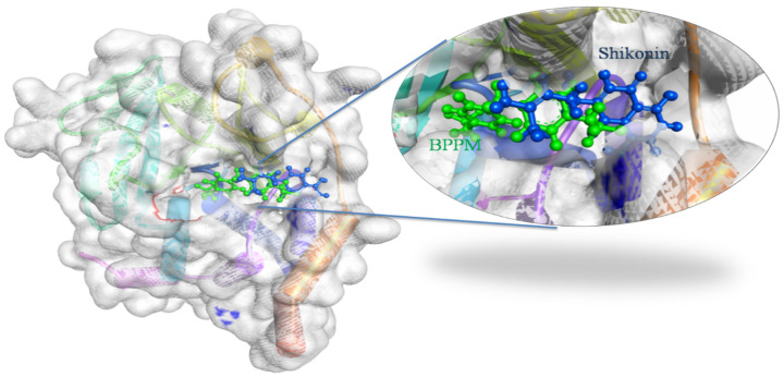
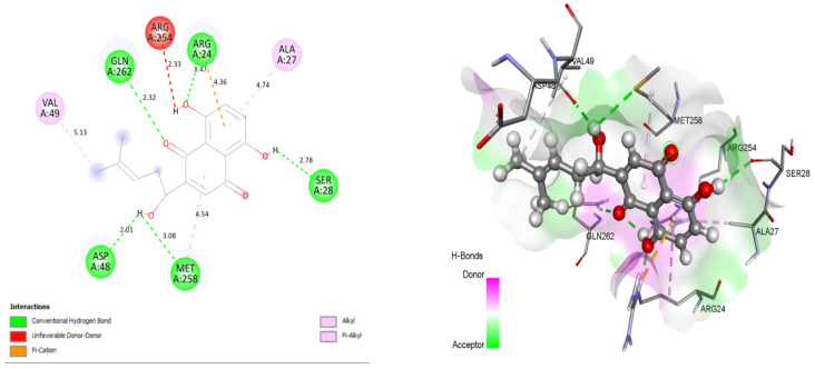
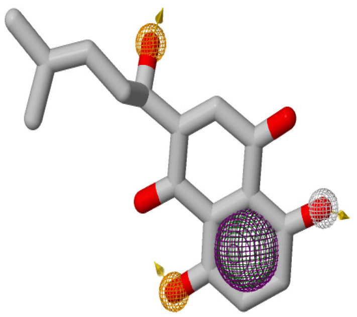
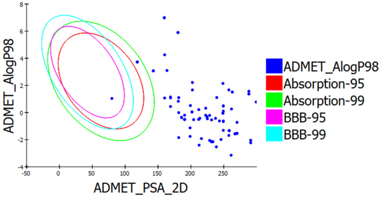
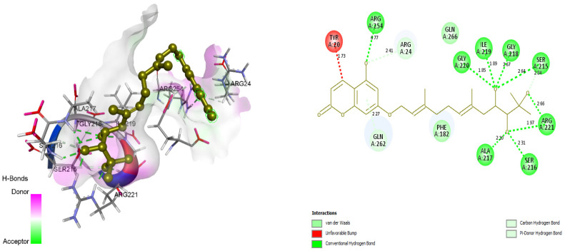
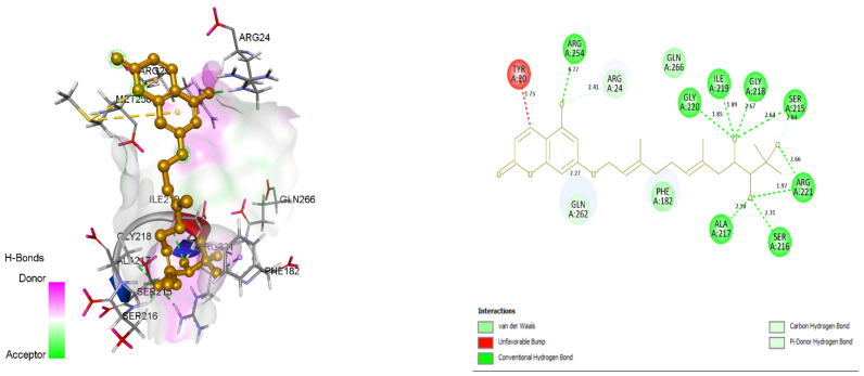
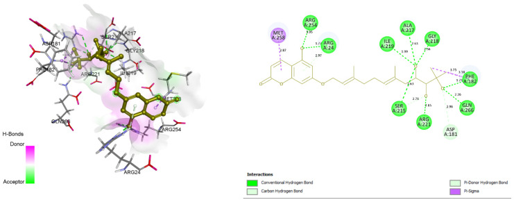
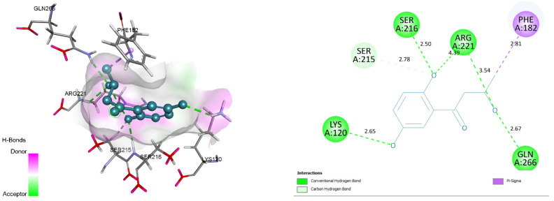
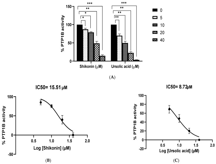
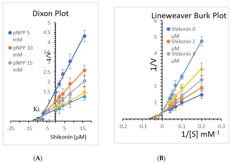
Diabetes mellitus is a multifactorial disease that affects both developing and developed countries and is a major public health concern. Many synthetic drugs are available in the market, which counteracts the associated pathologies. However, due to the propensity of side effects, there is an unmet need for the investigation of safe and effective drugs. This research aims to find a novel phytoconstituent having diminished action on blood glucose levels with the least side effects. Shikonin is a naturally occurring naphthoquinone dying pigment obtained by the roots of the Boraginaceae family. Besides its use as pigments, it can be used as an antimicrobial, anti-inflammatory, and anti-tumor agent. This research aimed to hypothesize the physicochemical and phytochemical properties of Shikonin's in silico interaction with protein tyrosine phosphate 1B, as well as it's in vitro studies, in order to determine its potential anti-diabetic impact. To do so, molecular docking experiments with target proteins were conducted to assess their anti-diabetic ability. Analyzing associations with corresponding amino acids revealed the significant molecular interactions between Shikonin and diabetes-related target proteins. In silico pharmacokinetics and toxicity profile of Shikonin using ADMET Descriptor, Toxicity Prediction, and Calculate Molecular Properties tools from Biovia Discovery Studio v4.5. Filter by Lipinski and Veber Rule's module from Biovia Discovery Studio v4.5 was applied to assess the drug-likeness of Shikonin. The in vitro studies exposed that Shikonin shows an inhibitory potential against the PTP1B with an IC50 value of 15.51 µM. The kinetics studies revealed that it has a competitive inhibitory effect (Ki = 7.5 M) on the enzyme system, which could be useful in the production of preventive and therapeutic agents. The findings of this research suggested that the Shikonin could be used as an anti-diabetic agent and can be used as a novel source for drug delivery.

Keywords: diabetes mellitus; hypoglycemic; molecular docking; protein-tyrosine phosphatase; shikonin.


Conflict of interest statement

The authors declare no conflict of interest.

Figures

Figure 1
Figure 1
Docking view of Shikonin in the binding sites of PTP1B (PDB ID: 1AAX). On the left side is the surface view of the docked complex-Shikonin and 1AAX, on the right side, a binding pocket is magnified to present Shikonin (shown in the blue-colored ball and stick view) superimposed on the co-crystal ligand-BPPM (shown in the green-colored ball and stick view). Diagrams are prepared in the Biovia Discovery Studio.
Figure 2
Figure 2
Docking view of Shikonin in the binding sites of PTP1B (PDB ID: 1AAX). On the left side is the stereo view of the docked complex-Shikonin and 1AAX, and on the right side are Shikonin-1AAX interactions presented in 2D. Diagrams are drawn in Discovery Studio, with hydrogenbonds shown as agreen dashed line. Protein structure residues are annotated with a 3-letter amino acid code, while Shikonin is presented in a ball-and-stick style.
Figure 3
Figure 3
Pharmacophore features generated for Shikonin on ZINCPharmer. The hydrophobic, hydrogen bond donor, and hydrogen bond acceptors, and aromatic features are displayed in mesh spheres of green, white, orange, and purple, respectively. The orange arrows indicate the constraint direction.
Figure 4
Figure 4
ALogP versus polar surface area (PSA) plot for Shikonin pharmacophores showing the 95% and 99% confidence limit ellipse corresponding to the blood-brain barrier (BBB) and intestinal absorption.
Figure 5
Figure 5
Docking view of ZINC31168041in the binding sites of PTP1B (PDB ID: 1AAX). On the left side is the stereo view of the docked complex-ZINC31168041 and 1AAX, on the right side, ZINC31168041-1AAX interactions are presented in 2D view. Diagrams are drawn in Discovery Studio, hydrogen-bonds are shown as agreen-dashed line along with the distance in Å. Protein structure residues are annotated with a 3-letter amino acid code, while ZINC31168041 is presented in agreen-colored ball and stick style.
Figure 6
Figure 6
Docking view of ZINC31168045 in the binding sites of PTP1B (PDB ID: 1AAX). On the left side is the stereo view of the docked complex-ZINC31168045 and 1AAX, on the right side ZINC31168045 -1AAX interactions are presented in 2D. Diagrams are drawn in Discovery Studio, hydrogen-bonds are shown in a green-dashed line along with the distance in Å. Protein structure residues are annotated with a 3-letter amino acid code, while ZINC31168045 is presented in anorange-colored ball and stick style.
Figure 7
Figure 7
Docking view of ZINC31168048 in the binding sites of PTP1B (PDB ID: 1AAX). On the left side is the stereo view of the docked complex-ZINC31168048 and 1AAX, and on the right side ZINC31168048 -1AAX interactions are presented in 2D. Diagrams are drawn in Discovery Studio, hydrogen-bonds are shown as a green-dashed line along with the distance in Å. Protein structure residues are annotated with a 3-letter amino acid code, while ZINC31168048 is presented in agreen-colored ball and stick style.
Figure 8
Figure 8
Docking view of ZINC31168554 in the binding sites of PTP1B (PDB ID: 1AAX). On theleft side is the stereo view of the docked complex- ZINC31168554 and 1AAX, on the right side ZINC31168554 -1AAX interactions are presented in 2D. Diagrams are drawn in Discovery Studio, hydrogen-bonds are shown as a green-dashed line along with the distance in Å. Protein structure residues are annotated with a 3-letter amino acid code, while ZINC31168554 is presented in ablue-colored ball and stick style.
Figure 9
Figure 9
Root means square deviation (RMSD) of protein and ligand after the initial RMSD values were stabilized. This plot shows RMSD values for protein on the left Y-axis, whereas for ligand, these values are indicated on the right Y-axis. The RMSD graph for the backbone is shown in green, and for theligand fit on protein in red.
Figure 10
Figure 10
The RMSF protein backbone and ligand complex, the red color shows the B factor, which means the PDB and green shows the interaction of the ligand to the protein.
Figure 11
Figure 11
Protein-ligand complex interaction during the molecular docking simulation.
Figure 12
Figure 12
(A) Dose-dependent protein tyrosine phosphatases 1B inhibitory activity of Shikonin. Ursolic acid, a strong inhibitor of PTP1B, was used as a positive control. The data shown are mean ± S.E.M. of three independent experiments performed in duplicate (* p < 0.01, ** p < 0.001, and *** p < 0.0001 represent a significant difference compared with the control). (B) IC50 graph of Shikonin against PTP1B. (C) IC50 graph of ursolic acid against PTP1B.
Figure 13
Figure 13
(A) Dixon plots of protein tyrosine phosphatases 1B (PTP1B) inhibition by Shikonin at various substrate (pNPP) concentrations (5, 10, 15, 20, and 25 mM). (B) Lineweaver–Burk plot for inhibition of PTP1B by Shikonin was analyzed in the presence of different concentrations of Shikonin (0, 2, 4, 8, and 16 µM).

References

    1. Shoaib A., Salem-Bekhit M.M., Siddiqui H.H., Dixit R.K., Bayomi M., Khalid M., Badruddeen, Shakeel F. Antidiabetic activity of standardized dried tubers extract of Aconitum napellus in streptozotocin-induced diabetic rats. 3 Biotech. 2020;10:56. doi: 10.1007/s13205-019-2043-7. - DOI - PMC - PubMed
    1. Rowley W.R., Bezold C. Creating public awareness: State 2025 diabetes forecasts. Popul. Health Manag. 2012;15:194–200. doi: 10.1089/pop.2011.0053. - DOI - PubMed
    1. Shaw J.E., Sicree R.A., Zimmet P.Z. Global estimates of the prevalence of diabetes for 2010 and 2030. Diabetes Res. Clin. Pract. 2010;87:4–14. doi: 10.1016/j.diabres.2009.10.007. - DOI - PubMed
    1. Al Dawish M.A., Robert A.A., Braham R., Al Hayek A.A., Al Saeed A., Ahmed R.A., Al Sabaan F.S. Diabetes Mellitus in Saudi Arabia: A Review of the Recent Literature. Curr. Diabetes Rev. 2016;12:359–368. doi: 10.2174/1573399811666150724095130. - DOI - PubMed
    1. Robert A.A., Al-Dawish A., Mujammami M., Dawish M.A.A. Type 1 Diabetes Mellitus in Saudi Arabia: A Soaring Epidemic. Int. J. Pediatr. 2018;2018:9408370. doi: 10.1155/2018/9408370. - DOI - PMC - PubMed

MeSH terms