. 2021 Jun 30;43(2):529-542.
doi: 10.3390/cimb43020040.

Analysis of Dip2B Expression in Adult Mouse Tissues Using the LacZ Reporter Gene

Affiliations

Analysis of Dip2B Expression in Adult Mouse Tissues Using the LacZ Reporter Gene

Rajiv Kumar Sah et al. Curr Issues Mol Biol. .

Abstract

Disconnected (disco)-interacting protein 2 homolog B (Dip2B) is a member of the Dip2 superfamily and plays an essential role in axonal outgrowth during embryogenesis. In adults, Dip2B is highly expressed in different brain regions, as shown by in situ analysis, and may have a role in axon guidance. However, the expression and biological role of Dip2B in other somatic tissues remain unknown. To better visualize Dip2B expression and to provide insight into the roles of Dip2B during postnatal development, we used a Dip2btm1a(wtsi)komp knock-in mouse model, in which a LacZ-Neo fusion protein is expressed under Dip2b promoter and allowed Dip2B expression to be analyzed by X-gal staining. qPCR analyses showed that Dip2b mRNA was expressed in a variety of somatic tissues, including lung and kidney, in addition to brain. LacZ staining indicated that Dip2B is broadly expressed in neuronal, reproductive, and vascular tissues as well as in the kidneys, heart, liver, and lungs. Moreover, neurons and epithelial cells showed rich staining. The broad and intense patterns of Dip2B expression in adult mice provide evidence of the distribution of Dip2B in multiple locations and, thereby, its implication in numerous physiological roles.

Keywords: Dip2b; LacZ; nervous system; reproductive system; respiratory system; vascular system.


Conflict of interest statement

The authors declare no conflict of interest.

Figures

Figure 1
Figure 1
Tm1a knockout-first reporter tagged insertion allele and genotyping. (A) Structure of Dip2btm1a (KOMP) Wtsi allele. A critical exon is flanked by loxP sites, with FRT, lacZ, neo, and FRT elements upstream of the critical exon. (B) Genotyping PCR results for tail biopsy from Dip2btm1a/+ intercrossing mice. A 300 bp band appeared on the gel for the transgene allele and a 350 bp band for the wild-type allele.
Figure 2
Figure 2
LacZ expression in the nervous system. (AF) Frozen section expression analysis of Dip2btm1a/+ brain regions at high magnification. (A) LacZ protein expression is notable in cortex neurons, (B) the hippocampus and dentate gyrus, (C) the granular and Purkinje cell layer of the cerebellum, (D) the ventral striatum, (E) the olfactory nucleus, and (F) the inner plexiform and outer nucleus layer of the olfactory bulb in Dip2btm1a/+. (GJ) LacZ staining of the adult spinal cord. (G) Whole-mount staining of the adult spinal cord. (H) Longitudinal section of the spinal cord depicting LacZ signals in the gray matter and central canal in Dip2btm1a/+. (I,J) The wild-type whole-mounted and frozen sections were LacZ-negative. (KP) LacZ staining in the adult eye. (K) Whole-mount staining of P56 mouse eyes, along with nerve fibers, showed LacZ staining. (L) Sagittal section of the eye stained for LacZ expression. The retinal wall and cornea showed LacZ expression in Dip2btm1a/+. (M) LacZ-positive retina of Dip2btm1a/+ at high magnification showed strong signals in the external limiting membrane, outer nuclear layer, and optic nerve fiber layer. (NP) Wild-type control was devoid of a LacZ-positive signal.
Figure 3
Figure 3
LacZ staining of the male reproductive system. (AF) Whole-mount LacZ-stained male testis, epididymis, seminal vesicle, and prostate gland. Non-specific staining can be seen in the wild-type epididymis and Dip2btm1a/+ seminal vesicle. (GN) Frozen section of a LacZ-stained male testis, epididymis, penis, and prostate gland. CuEp, cuboidal epithelium; CoEp, columnar epithelium; PrEp, perpetual epithelium; EpGi, glans epithelium.
Figure 4
Figure 4
LacZ expression in the female reproductive system. (A) Cross-section of the ovary of a non-pregnant mouse. LacZ expression is visible in the ovarian follicles and oviduct. (B,C) High magnification of ovarian follicles and oviduct. (D) Section of the vagina with a LacZ signal at the stratified squamous epithelium (SqEp). (EH) Wild-type littermate was LacZ-negative.
Figure 5
Figure 5
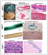
LacZ expression in the digestive system. Whole-mount and frozen section showing LacZ signals in the squamous epithelium (SqEp) of the fore stomach (AC), esophagus (G,H), crypts of the Lieberkühn of the jejunum (K), and dorsal epithelium of the tongue (L). The control, labeled as WT, shows no staining (DF,I,J).
Figure 6
Figure 6
LacZ expression in the respiratory system. (A) Whole-mount staining of the lung. (B) Coronal section of the lung, showing an X-gal signal in the epithelial lining of the bronchioles. (C) X-gal signals in the respiratory epithelium lining of the trachea and squamous epithelium of the larynx (D).
Figure 7
Figure 7
LacZ expression in the genitourinary system. (AC) Whole-mount and frozen section LacZ staining of a kidney, showing X-gal signal at the renal pelvis and glomeruli. (D) Frozen section of a urinary bladder, showing LacZ signals in the epithelium. (E) Frozen section of an adrenal gland, showing LacZ staining in the medulla.
Figure 8
Figure 8
LacZ expression in the cardiovascular system. (AD) Whole-mount LacZ staining of the veins collected from the tail, tongue, skin, and esophagus. (EG) LacZ staining in the heart, showing uniform staining at the atrium and ventricle myocardium.
Figure 9
Figure 9
qPCR results for total RNA of 8-week-old tissues. (A) Relative mRNA levels were calculated using the ddCt method. Data shown are representative of at least three independent experiments and expressed as the mean ± SD. (B) Confirmation of the specificity of product produced by qPCR using gel electrophoresis.

References

    1. Mukhopadhyay M., Pelka P., DeSousa D., Kablar B., Schindler A., Rudnicki M.A., Campos A.R. Cloning, Genomic Organization and Expression Pattern of a Novel Drosophila Gene, the Disco-Interacting Protein 2 (Dip2), and Its Murine Homolog. Gene. 2002;293:59–65. doi: 10.1016/S0378-1119(02)00694-7. - DOI - PubMed
    1. Ma J., Chen L., He X.-X., Wang Y.-J., Yu H.-L., He Z.-X., Zhang L.-Q., Zheng Y.-W., Zhu X.-J. Functional Prediction and Characterization of Dip2 Gene in Mice. Cell Biol. Int. 2019;43:421–428. doi: 10.1002/cbin.11106. - DOI - PubMed
    1. Winnepenninckx B., Debacker K., Ramsay J., Smeets D., Smits A., FitzPatrick D.R., Kooy R.F. CGG-Repeat Expansion in the DIP2B Gene Is Associated with the Fragile Site FRA12A on Chromosome 12q13.1. Am. J. Hum. Genet. 2007;80:221–231. doi: 10.1086/510800. - DOI - PMC - PubMed
    1. Campos A.R., Lee K.J., Steller H. Establishment of Neuronal Connectivity during Development of the Drosophila Larval Visual System. J. Neurobiol. 1995;28:313–329. doi: 10.1002/neu.480280305. - DOI - PubMed
    1. Heilig J.S., Freeman M., Laverty T., Lee K.J., Campos A.R., Rubin G.M., Steller H. Isolation and Characterization of the Disconnected Gene of Drosophila Melanogaster. EMBO J. 1991;10:809–815. doi: 10.1002/j.1460-2075.1991.tb08013.x. - DOI - PMC - PubMed