. 2021 Jun 30;13(7):2269.
doi: 10.3390/nu13072269.

Oral Ferric Maltol Does Not Adversely Affect the Intestinal Microbiome of Patients or Mice, But Ferrous Sulphate Does

Affiliations

Oral Ferric Maltol Does Not Adversely Affect the Intestinal Microbiome of Patients or Mice, But Ferrous Sulphate Does

Awad Mahalhal et al. Nutrients. .

Abstract

Background and aims: Altering dietary ferrous sulphate (FS) consumption exacerbates a murine model of colitis and alters the intestinal microbiome. We investigated the impact of oral ferric maltol (FM) and FS on mice with dextran sodium sulphate (DSS) induced colitis, and the microbiome of patients with iron deficiency.

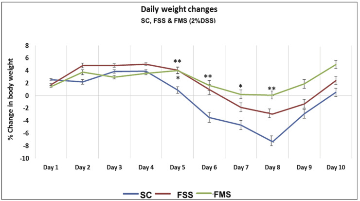
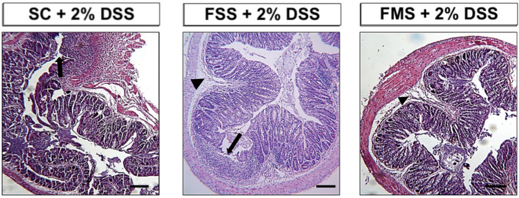
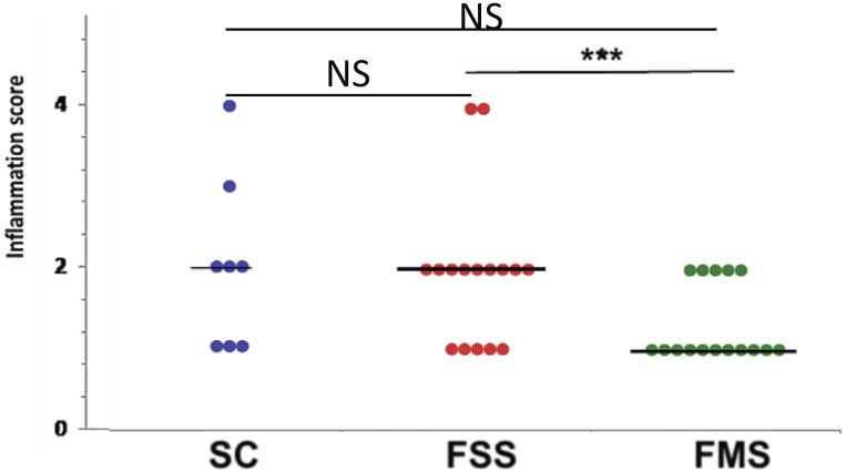
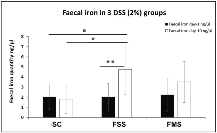
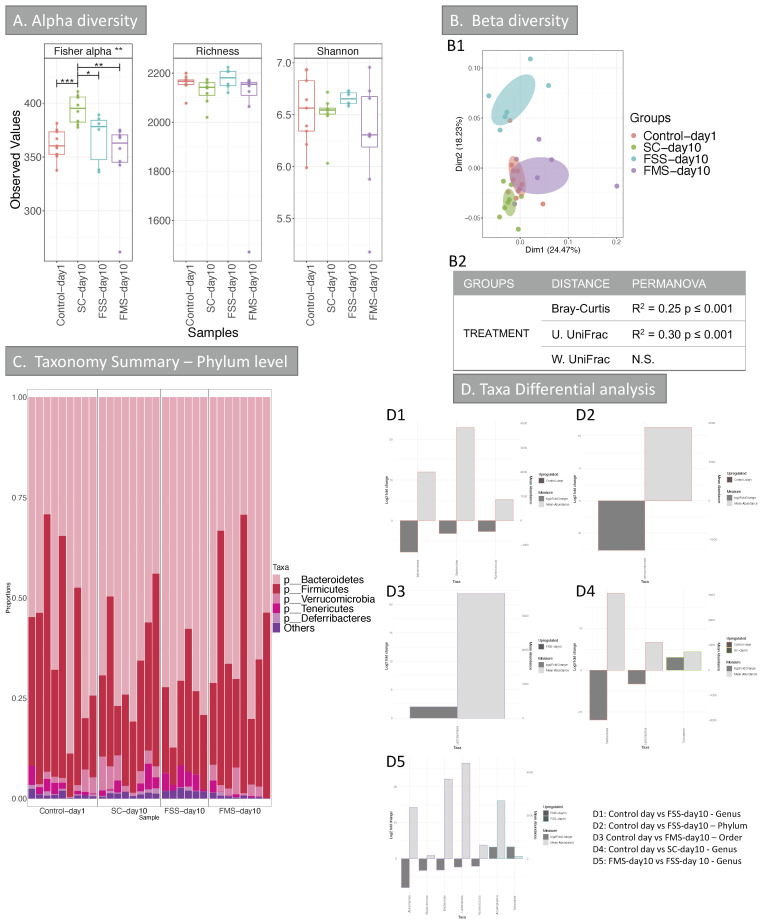
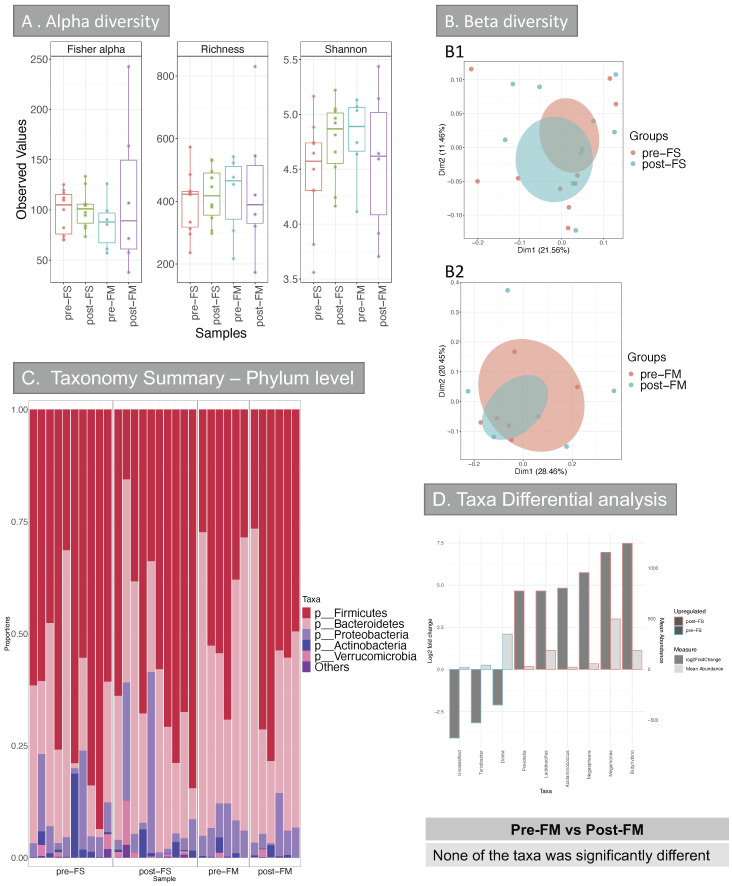
Methods: Mice had acute colitis induced, with 2% DSS for 5 days, followed by water. During this period, groups of mice were fed standard chow (200 ppm iron, SC, n = 8), or SC with 200ppm FS supplementation (n = 16, FSS), or SC with 200 ppm FM supplementation (n = 16, FMS). Clinical, pathological and microbiome assessments were compared at days 1 and 10. Fecal bacterial gDNA was extracted and the microbiome assessed by sequencing. Statistical inferences were made using MacQIIME. Principal Coordinates Analysis were used to visualize beta-diversity cluster analysis. Ten patients with IDA were treated with FS, and six with inactive inflammatory bowel disease received FM, supplements for four weeks: pre- and mid-treatment fecal samples were collected: the microbiome was assessed (see above).

Results: In mice, after DSS treatment, there was a decrease in many genera in the SC and FSS groups: Lactobacillales increased in mice that received FMS. In humans, FS treatment led to an increase in five genera, but FM was not associated with any measurable change. The severity of DSS-induced colitis was greater with FSS than FMS.

Conclusions: This study demonstrates differential and unique influences of ferric maltol and ferrous sulphate supplements on intestinal microbiota. These differences might contribute to the different side effects associated with these preparations.

Keywords: dysbiosis; iron; microbiome.


Conflict of interest statement

The authors declare no conflict of interest.

Figures

Figure 1
Figure 1
Percentage of weight change associated with acute DSS in mice receiving standard chow (SC) and chow supplemented with ferrous sulphate (FSS) or ferric maltol (FMS). Data are presented as a mean ± standard error of the mean. Statistical differences were assessed by Kruskal–Wallis test followed by Dunn’s multiple comparison tests (* p < 0.05, ** p < 0.01). (n = 40 ♀ mice) (asterisks above FMS vs. SC, asterisk below FSS vs. SC).
Figure 2
Figure 2
Representative haematoxylin- and eosin-stained sections of the distal colon from mice treated with 2% w/v dextran sodium sulphate for 5 days followed by another 5 days on plain drinking water. Arrowheads highlight submucosal oedema; arrows highlight almost complete loss of colonic epithelium. Scale Bar: 200 µm.
Figure 3
Figure 3
Spread plot of inflammation (colitis) scores for each group of mice at necropsy, at day 10. Horizontal lines at the median. Differences tested by one-way ANOVA (overall p = 0.06) followed by multiple comparisons Dunn’s test (*** p < 0.001). (NS non-significant).
Figure 4
Figure 4
Faecal iron concentration at two different time points (day 1 and 10) for three DSS-treated groups of mice (SC, FSS and FMS diets). Data are presented as a mean ± standard error of the mean. Differences were tested by t-test (inter-comparison) and by one-way ANOVA (intra-comparison) followed by post hoc test. (* p < 0.05, ** p < 0.01).
Figure 5
Figure 5
Alpha and Beta diversity, and taxonomy results of the intestinal microbiome (mice experiment). (A) Alpha diversity (OTUs level) of the intestinal microbiome from the 4 groups: Control-day 1, SC-day 10, FSS-day 10 and FMS-day 10. Three indices were considered: Fisher alpha (a parametric index that models species’ abundance as logseries distribution), richness (number of species) and Shannon index (a widely used index that considers species’ abundance and evenness). Pair-wise ANOVA was calculated between the groups and if significant, stars are shown on top (* p < 0.05, ** p < 0.01 and *** p < 0.001). (B) Beta diversity results; (B1) Principal Co-ordinate Analysis (PCOA) showing clustering of samples. The chart was produced using unweighted UniFrac (UniFrac) at OTUs level. Ellipses are 95% confidence interval of standard error. The table in (B2) summarises PERMANOVA results for all the distances, Bray-Curtis, unweighted UniFrac (U. UniFrac) and weighted (W. UniFrac). R2 refers to the percentage of variability among samples’ microbiome that can be explained by that factor/metadata. (C) Taxonomy summary for stool at phylum level. (D) Taxa differential analysis at phylum, order and genus levels are presented through bar charts; these show Log2 fold change between the groups compared, (y axis on the left and dark grey bar) and the mean abundance across all the samples (y axis on the right and light grey bar), details of the comparisons per chart are in the right bottom of the chart. Detail of taxa differential analysis results, including p values and adjusted p values, is in Supplementary Table S3. FS = ferrous sulphate and FM = ferric maltol.
Figure 6
Figure 6
Alpha and Beta diversity, and taxonomy results of the intestinal microbiome (Human cohort). (A) Alpha diversity (OTUs level) of the intestinal microbiome from the 4 groups: pre-FS, post-FS, pre-FM, post-FM. Three indices were considered: Fisher alpha (a parametric index that models species’ abundance as logseries distribution), richness (number of species) and Shannon index (a widely used index that considers species’ abundance and evenness). Pair-wise ANOVA was calculated between the groups, but none was significant. (B) Beta diversity results (B1/B2) Principal Co-ordinate Analysis (PCOA) showing c––lustering of samples. The chart was produced using unweighted UniFrac (UniFrac) at OTUs level. Ellipses are 95% confidence interval of standard error. PERMANOVA analysis comparing the groups for all the distances (Bray-Curtis, unweighted UniFrac (UniFrac) and weighted (W. UniFrac)) were not significant. (C) Taxonomy summary at phylum level. (D) Taxa differential analysis at genus level are presented through bar charts, which show Log2 fold change between pre-FS vs. post-FS samples, (y axis on the left and dark grey bar) and the mean abundance across all the samples (y axis on the right and light grey bar). The comparison of samples from patients that were given FM before and after treatment (pre-FM vs. post-FM) did not give any significant results at any of the taxonomical levels analysed (phylum, class, order, family, genus and OTUs). Detail of taxa differential analysis results, including p values and adjusted p values, is in Supplementary Table S3. FS = ferrous sulphate and FM = ferric maltol.

Similar articles

Cited by

References

    1. Babbs C.F. Oxygen radicals in ulcrative colitis. Free Radic. Biol. Med. 1992;13:169–181. doi: 10.1016/0891-5849(92)90079-V. - DOI - PubMed
    1. Lund E.K., Wharf S.G., Fairweather-Tait S.J., Johnson I. Oral ferrous sulfate supplements increase the free radical-generating capacity of feces from healthy volunteers. Am. J. Clin. Nutr. 1999;69:250–255. doi: 10.1093/ajcn/69.2.250. - DOI - PubMed
    1. De Silva A.D., Tsironi E., Feakins R.M., Rampton D.S. Efficacy and tolerability of oral iron therapy in inflammatory bowel disease: A prospective, comparative trial. Aliment. Pharmacol. Ther. 2005;22:1097–1105. doi: 10.1111/j.1365-2036.2005.02700.x. - DOI - PubMed
    1. Xu S., He Y., Lin L., Chen P., Chen M., Zhang S. The emerging role of ferroptosis in intestinal disease. Cell Death Dis. 2021;12:289. doi: 10.1038/s41419-021-03559-1. - DOI - PMC - PubMed
    1. Stein J., Bager P., Befrits R., Gasche C., Gudehus M., Lerebours E., Magro F., Mearin F., Mitchell D., Oldenburg B., et al. Anaemia management in patients with inflammatory bowel disease: Routine practice across nine European countries. Eur. J. Gastroenterol. Hepatol. 2013;25:1456–1463. doi: 10.1097/MEG.0b013e328365ca7f. - DOI - PubMed

MeSH terms

LinkOut - more resources