Review
. 2021 Jun 29;22(13):6986.
doi: 10.3390/ijms22136986.

The Role of Cellular Stress in Intrauterine Growth Restriction and Postnatal Dysmetabolism

Affiliations
Review

The Role of Cellular Stress in Intrauterine Growth Restriction and Postnatal Dysmetabolism

Shelby L Oke et al. Int J Mol Sci. .

Abstract

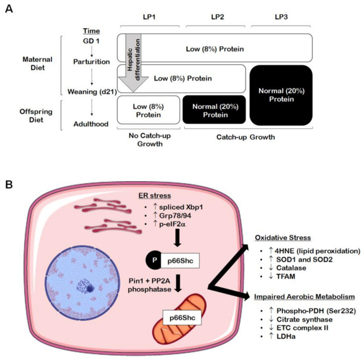
Disruption of the in utero environment can have dire consequences on fetal growth and development. Intrauterine growth restriction (IUGR) is a pathological condition by which the fetus deviates from its expected growth trajectory, resulting in low birth weight and impaired organ function. The developmental origins of health and disease (DOHaD) postulates that IUGR has lifelong consequences on offspring well-being, as human studies have established an inverse relationship between birth weight and long-term metabolic health. While these trends are apparent in epidemiological data, animal studies have been essential in defining the molecular mechanisms that contribute to this relationship. One such mechanism is cellular stress, a prominent underlying cause of the metabolic syndrome. As such, this review considers the role of oxidative stress, mitochondrial dysfunction, endoplasmic reticulum (ER) stress, and inflammation in the pathogenesis of metabolic disease in IUGR offspring. In addition, we summarize how uncontrolled cellular stress can lead to programmed cell death within the metabolic organs of IUGR offspring.

Keywords: cell death; cell stress; intrauterine growth restriction (IUGR), metabolism; metabolic syndrome.


Conflict of interest statement

The authors declare no conflict of interest.

Figures

Figure 1
Figure 1
Perinatal maternal protein restriction (MPR) in combination with postnatal catch-up growth leads to endoplasmic reticulum (ER) stress, oxidative stress, and mitochondrial dysfunction in adult male rat offspring. (A) Percent protein composition and the timing of nutritional intervention differ across the various groups of MPR offspring ((A); LP1, LP2, LP3) in order to study the effects of postnatal catch-up growth on molecular outcomes in the growth-restricted liver. (B) Male offspring weaned onto a normal (20%) protein diet following the completion of hepatic differentiation (e.g., LP2 offspring) display increased markers of hepatic ER stress (i.e., increased spliced Xbp1, Grp78/94, and p-eIF2α) in adulthood, leading to elevation of the oxidative stress marker p66Shc. These same offspring further exhibit increased markers of oxidative stress (i.e., increased 4HNE and SOD1/2; decreased catalase and TFAM) and aberrant markers of aerobic metabolism (i.e., increased p-PDH[Ser232] and LDHa; decreased citrate synthase and complex II).
Figure 2
Figure 2
Maternal protein restriction (MPR) leads to increased hepatic lipid peroxidation in male rat offspring at four months of age. Representative western immunoblots illustrating expression of 4-hydroxynonenol (4HNE), a marker of lipid peroxidation, in (A) LP1, (B), LP2, and (C) LP3 offspring relative to control offspring. (D) 4HNE abundance for each group was compared against that of control offspring and analyzed using a two-tailed unpaired Student’s t-test. 4HNE abundance was expressed as means normalized to total protein abundance ± SEM (n = 7–8/group). ** Significant difference (p < 0.01), *** significant difference (p < 0.001).

References

    1. Ravelli G.P., Stein Z.A., Susser M.W. Obesity in Young Men after Famine Exposure in Utero and Early Infancy. N. Engl. J. Med. 1976;295:349–353. doi: 10.1056/NEJM197608122950701. - DOI - PubMed
    1. Ravelli A.C., van der Meulen J.H., Michels R.P., Osmond C., Barker D.J., Hales C.N., Bleker O.P. Glucose Tolerance in Adults after Prenatal Exposure to Famine. Lancet. 1998;351:173–177. doi: 10.1016/S0140-6736(97)07244-9. - DOI - PubMed
    1. Ravelli A.C., van der Meulen J.H., Osmond C., Barker D.J., Bleker O.P. Obesity at the Age of 50 y in Men and Women Exposed to Famine Prenatally. Am. J. Clin. Nutr. 1999;70:811–816. doi: 10.1093/ajcn/70.5.811. - DOI - PubMed
    1. Baschat A.A. Pathophysiology of Fetal Growth Restriction: Implications for Diagnosis and Surveillance. Obstet. Gynecol. Surv. 2004;59:617–627. doi: 10.1097/01.OGX.0000133943.54530.76. - DOI - PubMed
    1. Sharma D., Shastri S., Sharma P. Intrauterine Growth Restriction: Antenatal and Postnatal Aspects. Clin. Med. Insights. Pediatrics. 2016;10:67–83. doi: 10.4137/CMPed.S40070. - DOI - PMC - PubMed

MeSH terms