

Differences between Human and Mouse IgM Fc Receptor (FcµR)
- PMID: 34209905
- PMCID: PMC8267714
- DOI: 10.3390/ijms22137024
Differences between Human and Mouse IgM Fc Receptor (FcµR)
Abstract
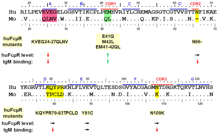
Both non-immune "natural" and antigen-induced "immune" IgM are important for protection against pathogens and for regulation of immune responses to self-antigens. Since the bona fide IgM Fc receptor (FcµR) was identified in humans by a functional cloning strategy in 2009, the roles of FcµR in these IgM effector functions have begun to be explored. In this short essay, we describe the differences between human and mouse FcµRs in terms of their identification processes, cellular distributions and ligand binding activities with emphasis on our recent findings from the mutational analysis of human FcµR. We have identified at least three sites of human FcµR, i.e., Asn66 in the CDR2, Lys79 to Arg83 in the DE loop and Asn109 in the CDR3, responsible for its constitutive IgM-ligand binding. Results of computational structural modeling analysis are consistent with these mutational data and a model of the ligand binding, Ig-like domain of human FcµR is proposed. Serendipitously, substitution of Glu41 and Met42 in the CDR1 of human FcµR with mouse equivalents Gln and Leu, either single or more prominently in combination, enhances both the receptor expression and IgM binding. These findings would help in the future development of preventive and therapeutic interventions targeting FcµR.
Keywords: 3D structure; FcR; FcµR; Fcα/µR; IgM binding; computational structural model; pIgR; species difference.
Conflict of interest statement
The authors have no conflict of interest.
Figures


Similar articles
-
Physiological and Pathophysiological Roles of IgM Fc Receptor (FcµR) Isoforms.Int J Mol Sci. 2023 Mar 17;24(6):5728. doi: 10.3390/ijms24065728. Int J Mol Sci. 2023. PMID: 36982860 Free PMC article. Review.
-
Identification of Amino Acid Residues in Human IgM Fc Receptor (FcµR) Critical for IgM Binding.Front Immunol. 2021 Jan 27;11:618327. doi: 10.3389/fimmu.2020.618327. eCollection 2020. Front Immunol. 2021. PMID: 33584711 Free PMC article.
-
The three complementarity-determining region-like loops in the second extracellular domain of human Fc alpha/mu receptor contribute to its binding of IgA and IgM.Immunobiology. 2013 May;218(5):798-809. doi: 10.1016/j.imbio.2012.09.004. Epub 2012 Oct 4. Immunobiology. 2013. PMID: 23182711
-
Functions of IgM fc receptor (FcµR) related to autoimmunity.Autoimmunity. 2024 Dec;57(1):2323563. doi: 10.1080/08916934.2024.2323563. Epub 2024 Mar 11. Autoimmunity. 2024. PMID: 38465789 Review.
-
The Appearance and Diversification of Receptors for IgM During Vertebrate Evolution.Curr Top Microbiol Immunol. 2017;408:1-23. doi: 10.1007/82_2017_22. Curr Top Microbiol Immunol. 2017. PMID: 28884191 Review.
Cited by
-
My Name Is Legion, for We Are Many-The Complex Community of Antibody Receptors.Int J Mol Sci. 2023 Oct 16;24(20):15226. doi: 10.3390/ijms242015226. Int J Mol Sci. 2023. PMID: 37894907 Free PMC article.
-
An Unexpected Role for Cell Density Rather than IgM in Cell-Surface Display of the Fc Receptor for IgM on Human Lymphocytes.Immunohorizons. 2022 Jan 18;6(1):47-63. doi: 10.4049/immunohorizons.2100094. Immunohorizons. 2022. PMID: 35042773 Free PMC article.
-
Structural basis for Fc receptor recognition of immunoglobulin M.Nat Struct Mol Biol. 2023 Jul;30(7):1033-1039. doi: 10.1038/s41594-023-00985-x. Epub 2023 Apr 24. Nat Struct Mol Biol. 2023. PMID: 37095205 Free PMC article.
-
Physiological and Pathophysiological Roles of IgM Fc Receptor (FcµR) Isoforms.Int J Mol Sci. 2023 Mar 17;24(6):5728. doi: 10.3390/ijms24065728. Int J Mol Sci. 2023. PMID: 36982860 Free PMC article. Review.
-
Immunoglobulin M perception by FcμR.Nature. 2023 Mar;615(7954):907-912. doi: 10.1038/s41586-023-05835-w. Epub 2023 Mar 22. Nature. 2023. PMID: 36949194
References
Publication types
MeSH terms
Substances
LinkOut - more resources
Full Text Sources
Molecular Biology Databases
Miscellaneous
