Review
. 2021 Jun 29;22(13):7030.
doi: 10.3390/ijms22137030.

Evidence for the Role of Mitochondrial DNA Release in the Inflammatory Response in Neurological Disorders

Affiliations
Review

Evidence for the Role of Mitochondrial DNA Release in the Inflammatory Response in Neurological Disorders

Gonzalo E Moya et al. Int J Mol Sci. .

Abstract

Mitochondria are regarded as the metabolic centers of cells and are integral in many other cell processes, including the immune response. Each mitochondrion contains numerous copies of mitochondrial DNA (mtDNA), a small, circular, and bacterial-like DNA. In response to cellular damage or stress, mtDNA can be released from the mitochondrion and trigger immune and inflammatory responses. mtDNA release into the cytosol or bloodstream can occur as a response to hypoxia, sepsis, traumatic injury, excitatory cytotoxicity, or drastic mitochondrial membrane potential changes, some of which are hallmarks of neurodegenerative and mood disorders. Released mtDNA can mediate inflammatory responses observed in many neurological and mood disorders by driving the expression of inflammatory cytokines and the interferon response system. The current understanding of the role of mtDNA release in affective mood disorders and neurodegenerative diseases will be discussed.

Keywords: inflammation; mitochondria; mitochondrial DNA (mtDNA); neurodegenerative disease; neuropsychiatric disorder; reactive oxygen species (ROS).


Conflict of interest statement

P.D.R. is on the board of the Ottagan Addiction Recovery program. G.E.M. and K.E.D.-R. declare no conflict of interest.

Figures
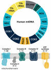
Figure 1
Figure 1
Overview of mtDNA. Mitochondrial DNA encodes for 13 protein subunits involved in oxidative phosphorylation (OXPHOS), the main energy-producing pathway of the cell. The diagram depicts the protein coding genes found within the mitochondrial genome that encode for subunits of OXPHOS complexes. Genes are color coded to correspond to the color of the OXPHOS complex. The numbers reflect the number of protein subunits of each complex encoded by the mitochondria out of the total number of protein subunits. The genetic instructions for the remaining subunits are carried in the nucleus. The 22 transfer RNAs encoded by the mitochondrial genome are not shown. Abbreviations: NADH dehydrogenase/Complex I subunits (ND1-6); Cytochrome oxidase/Complex III subunits (COX1-3); ATP synthase subunits (ATP6, ATP8); cytochrome b (CYTb).
Figure 2
Figure 2
The inflammatory response after mtDNA release from mitochondria or ccf-mtDNA internalization. (A) Internalization of ccf-mtDNA leads to (B) activation of Toll-like receptor 9 (TLR9) and (C) upregulation of nuclear factor-κB (NF-κB), which (D) promotes the expression of inflammatory cytokines. Additionally, a rise in intracellular reactive oxygen species (ROS) causes (E) NOD-, LRR-, and pyrin domain-containing protein 3 (NLRP3) activation, ultimately leading to NLRP3 inflammasome assembly. This causes caspase-1 (CAS1)-mediated pro-interleukin (pro-IL) activation, leading to either localized inflammation or cytokine release. Alternatively, (F) mtDNA can be released to the cytosol and bind cyclic GMP-AMP synthase (cGAS) following endosomal rupture, or (G) mtDNA release from mitochondria. mtDNA release from the mitochondria can be a result of mitochondrial dysfunction or impaired mitophagy. (H) cGAS-bound mtDNA can lead to a type I interferon (IFN) response, which further contributes to the inflammatory response from (E). While mtDNA is depicted in an intact and circular form, mtDNA fragments in linear or circular form may also elicit similar immune responses.
Figure 3
Figure 3
A schematic representation of mtDNA-mediated inflammation in relevant neurodegenerative diseases. The known alterations that lead to inflammation and neurodegenerative disease are presented. Different mutations or aberrancies lead to accumulation of damage in different ways. We propose that accumulation of defective mitochondrial due to impaired mitophagy and the accumulation of oxidative stress are common factors that link mtDNA-dependent inflammation within these pathologies. Abbreviations: amyloid-beta (Aβ); NOD-like receptor, pyrin containing protein 3 (NLRP3); reactive oxygen species (ROS); superoxide dismutase (SOD); TAR DNA-binding protein 43 (TDP-43); TANK-binding kinase 1 (TBK1).

References

    1. Protasoni M., Zeviani M. Mitochondrial Structure and Bioenergetics in Normal and Disease Conditions. Int. J. Mol. Sci. 2021;22:586. doi: 10.3390/ijms22020586. - DOI - PMC - PubMed
    1. Bestwick M.L., Shadel G.S. Accessorizing the human mitochondrial transcription machinery. Trends Biochem. Sci. 2013;38:283–291. doi: 10.1016/j.tibs.2013.03.006. - DOI - PMC - PubMed
    1. Raha S., Robinson B.H. Mitochondria, oxygen free radicals, disease and ageing. Trends Biochem. Sci. 2000;25:502–508. doi: 10.1016/S0968-0004(00)01674-1. - DOI - PubMed
    1. Kim J., Gupta R., Blanco L.P., Yang S., Shteinfer-Kuzmine A., Wang K., Kang H. VDAC oligomers form mitochondrial pores to release mtDNA fragments and promote lupus-like disease. Science. 2019;366:1531–1536. doi: 10.1126/science.aav4011. - DOI - PMC - PubMed
    1. Picca A., Calvani R., Coelho-Junior H.J., Landi F., Bernabei R., Marzetti E. Mitochondrial Dysfunction, Oxidative Stress, and Neuroinflammation: Intertwined Roads to Neurodegeneration. Antioxidants. 2020;9:647. doi: 10.3390/antiox9080647. - DOI - PMC - PubMed

Substances