. 2021 Jun 29;22(13):7002.
doi: 10.3390/ijms22137002.

Sex-Based Differences in Cardiac Gene Expression and Function in BDNF Val66Met Mice

Affiliations

Sex-Based Differences in Cardiac Gene Expression and Function in BDNF Val66Met Mice

Marcus Negron et al. Int J Mol Sci. .

Abstract

Brain-derived neurotrophic factor (BDNF) is a pleiotropic neuronal growth and survival factor that is indispensable in the brain, as well as in multiple other tissues and organs, including the cardiovascular system. In approximately 30% of the general population, BDNF harbors a nonsynonymous single nucleotide polymorphism that may be associated with cardiometabolic disorders, coronary artery disease, and Duchenne muscular dystrophy cardiomyopathy. We recently showed that transgenic mice with the human BDNF rs6265 polymorphism (Val66Met) exhibit altered cardiac function, and that cardiomyocytes isolated from these mice are also less contractile. To identify the underlying mechanisms involved, we compared cardiac function by echocardiography and performed deep sequencing of RNA extracted from whole hearts of all three genotypes (Val/Val, Val/Met, and Met/Met) of both male and female Val66Met mice. We found female-specific cardiac alterations in both heterozygous and homozygous carriers, including increased systolic (26.8%, p = 0.047) and diastolic diameters (14.9%, p = 0.022), increased systolic (57.9%, p = 0.039) and diastolic volumes (32.7%, p = 0.026), and increased stroke volume (25.9%, p = 0.033), with preserved ejection fraction and fractional shortening. Both males and females exhibited lower heart rates, but this change was more pronounced in female mice than in males. Consistent with phenotypic observations, the gene encoding SERCA2 (Atp2a2) was reduced in homozygous Met/Met mice but more profoundly in females compared to males. Enriched functions in females with the Met allele included cardiac hypertrophy in response to stress, with down-regulation of the gene encoding titin (Tcap) and upregulation of BNP (Nppb), in line with altered cardiac functional parameters. Homozygous male mice on the other hand exhibited an inflammatory profile characterized by interferon-γ (IFN-γ)-mediated Th1 immune responses. These results provide evidence for sex-based differences in how the BDNF polymorphism modifies cardiac physiology, including female-specific alterations of cardiac-specific transcripts and male-specific activation of inflammatory targets.

Keywords: Duchenne muscular dystrophy; Val66Met; brain-derived neurotrophic growth factor; dilated cardiomyopathy; rs6265 polymorphism.


Conflict of interest statement

The authors have no conflicts of interest to disclose.

Figures
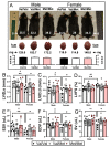
Figure 1
Figure 1
Val66Met leads to increased end systolic and diastolic volumes in female mice. (A) Representative 12-week-old littermate mice along with excised hearts from the same mice, showing greater body and heart weights in both males and females with the Met allele. Bars beneath representative hearts show similar heart weight-to-body weight ratios (HW/BW) among genotypes (n = 4 per group). (BG) Plot showing results of echocardiography assessment of littermate-matched male and female mice (8–12 weeks) with one (Val/Met, white bars and circles) or both (Met/Met, red bars and circles) of the rs6265 alleles, relative to non-carrier controls (light gray bars and black circles). Parameters shown (y axis) are (B) left ventricular internal dimension at end systole (LVID;s) and (C) diastole (LVID;d), (D) percent (%), fractional shortening (FS), (E) end systolic volume (ESV), (F) end diastolic volume (EDV), and (G) stroke volume (SR). n = 9 (Male Val/Val), n = 17 (Male Val/Met), n = 7 (Male Met/Met), n = 6 (Female Val/Val), n = 13 (Female Val/Met), n = 6 (Female Met/Met). Asterisks represent statistical significance between male and female Val/Val mice or between Val/Val versus Val/Met and Met/Met female mice. * p < 0.05and ** p < 0.001 using two-way ANOVA. # p < 0.05 and ## p < 0.01 using Mann–Whitney, ns = not significant.
Figure 2
Figure 2
The Met allele increases sex-based differences in global cardiac transcriptional profiles. Venn diagrams showing overlap of differentially expressed genes in whole hearts of male (A) and female (B) Val/Met versus Val/Val mice compared to those altered in Met/Met versus Val/Val mice. (C) Principal components analysis (PCA) shows unique whole heart male and female expression profiles in Val66Met mice. Groups are as indicated and colored as follows: purple = Male Val/Val, teal = Male Val/Met, green = Male Met/Met, red = Female Val/VaWe did not use any hyphensl, orange = Female Val/Met, blue = Female Met/Met. (D) Venn diagram showing overlap in differential genes for direct comparisons of males versus females for each genotype (green = Val/Val, blue = Val/Met, red = Met/Met). (EG), Volcano plots for males versus females for each genotype. The ordinate represents increasing significance, and the abscissa shows magnitude of change. Lines represent significance cutoffs, with numbers of genes significantly differentially expressed shown in parentheses. Red and blue dots represent significant genes more highly expressed in whole hearts of females and males, respectively. Grey dots represent genes not deemed as significantly different (p > 0.05 and/or fold < 1.5).
Figure 3
Figure 3
Met/Met mice exhibit a male-specific inflammatory profile. Bar chart shows enriched pathways (top bars), diseases and biofunctions (middle bars), and predicted upstream regulators (bottom) for whole hearts of male (left side) and female (right side) Met/Met mice compared to those of normal Val/Val controls. Bar size reflects significance (shown on x axis), and color represents Z score (significance of prediction of directionality), as indicated in the legend. Numbers in parentheses indicate numbers of genes in the listed category that were altered in hearts of either male or female Met/Met mice compared to respective controls.
Figure 4
Figure 4
Whole hearts from Val/Met mice exhibit blunted functional profiles loosely related to immune cell proliferation. Bar chart shows enriched pathways (top bars), diseases and biofunctions (middle bars), and predicted upstream regulators (bottom) for whole hearts of male (left side) and female (right side) Val/Met mice compared to normal Val/Val controls. Bar size reflects significance (shown on x axis), and color represents Z score (significance of prediction of directionality), as indicated in the legend. Numbers in parentheses indicate numbers of genes in the listed category that were altered in hearts of either male or female Val/Met mice compared to respective controls.
Figure 5
Figure 5
qPCR of select genes validates differences in whole hearts of males and females with and without the Met allele. Bar charts showing mRNA relative to GAPDH of titin-cap, Tcap (A), natriuretic peptide, Nppb (B), myosin light chain 2, Myl2 (C), and Atp2a2 (D) for males (black bars) and females (white bars) in each group. n = 3 or 4 each group, * p < 0.05 for pairwise comparisons between same-sex Val/Met or Met/Met versus respective Val/Val control mice. *** p = 0.0003 for pairwise comparison between male and female Met/Met mice. # p < 0.05 and ## p = 0.0007 using Mann–Whitney. Relative mRNA was calculated using the ΔΔ CT method with GAPDH as the internal normalization target and male Val/Val as the reference sample.

References

    1. Mowla S.J., Farhadi H.F., Pareek S., Atwal J.K., Morris S.J., Seidah N., Murphy R.A. Biosynthesis and Post-translational Processing of the Precursor to Brain-derived Neurotrophic Factor. J. Biol. Chem. 2001;276:12660–12666. doi: 10.1074/jbc.M008104200. - DOI - PubMed
    1. Teng K., Felice S., Kim T., Hempstead B.L. Understanding proneurotrophin actions: Recent advances and challenges. Dev. Neurobiol. 2010;70:350–359. doi: 10.1002/dneu.20768. - DOI - PMC - PubMed
    1. Roebroek A.J.M., Schalken J.A., Bussemakers M.J.G., van Heerikhuizen H., Onnekink C., Debruyne F.M.J., Bloemers H.P.J., Van de Ven W.J.M. Characterization of human c-fes/fps reveals a new transcription unit (fur) in the immediately upstream region of the proto-oncogene. Mol. Biol. Rep. 1986;11:117–125. doi: 10.1007/BF00364823. - DOI - PubMed
    1. Gray K., Ellis V. Activation of pro-BDNF by the pericellular serine protease plasmin. FEBS Lett. 2008;582:907–910. doi: 10.1016/j.febslet.2008.02.026. - DOI - PubMed
    1. Mizoguchi H., Nakade J., Tachibana M., Ibi D., Someya E., Koike H., Kamei H., Nabeshima T., Itohara S., Takuma K., et al. Matrix Metalloproteinase-9 Contributes to Kindled Seizure Development in Pentylenetetrazole-Treated Mice by Converting Pro-BDNF to Mature BDNF in the Hippocampus. J. Neurosci. 2011;31:12963–12971. doi: 10.1523/JNEUROSCI.3118-11.2011. - DOI - PMC - PubMed