

Family with sequence similarity 13 member A mediates TGF-β1-induced EMT in small airway epithelium of patients with chronic obstructive pulmonary disease
- PMID: 34210319
- PMCID: PMC8247231
- DOI: 10.1186/s12931-021-01783-z
Family with sequence similarity 13 member A mediates TGF-β1-induced EMT in small airway epithelium of patients with chronic obstructive pulmonary disease
Abstract
Background: To explore the role of family with sequence similarity 13 member A (FAM13A) in TGF-β1-induced EMT in the small airway epithelium of patients with chronic obstructive pulmonary disease (COPD).
Methods: Small airway wall thickness and protein levels of airway remodeling markers, EMT markers, TGF-β1, and FAM13A were measured in lung tissue samples from COPD and non-COPD patients. The correlations of FAM13A expression with COPD severity and EMT marker expression were evaluated. Gain- and loss-of-function assays were performed to explore the functions of FAM13A in cell proliferation, motility, and TGF-β1-induced EMT marker alterations in human bronchial epithelial cell line BEAS-2B.
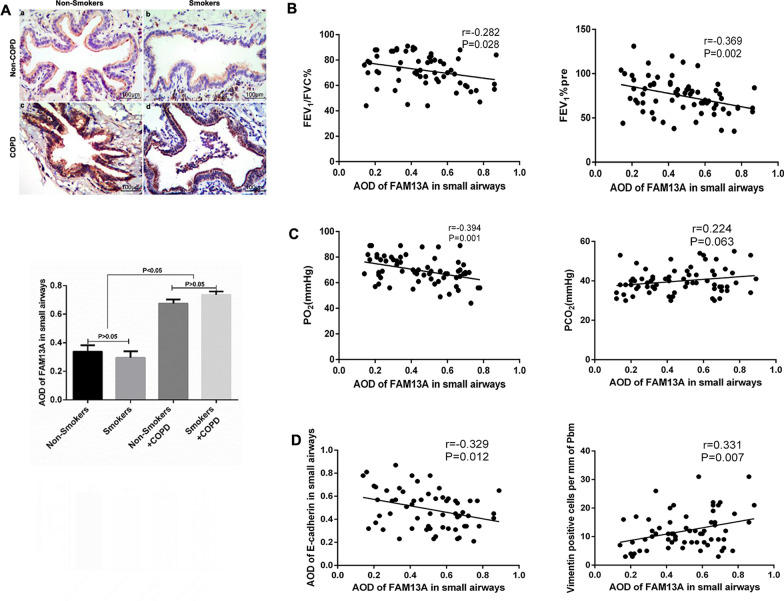
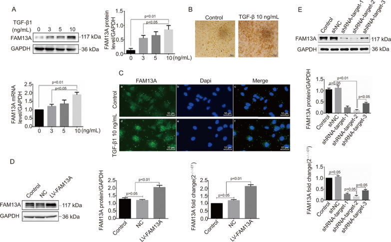
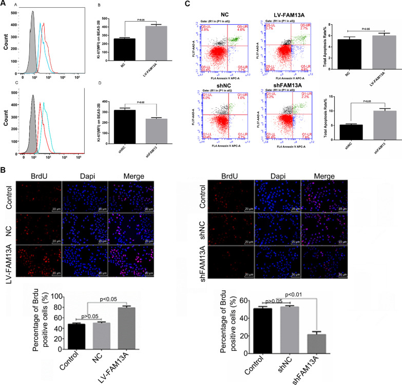
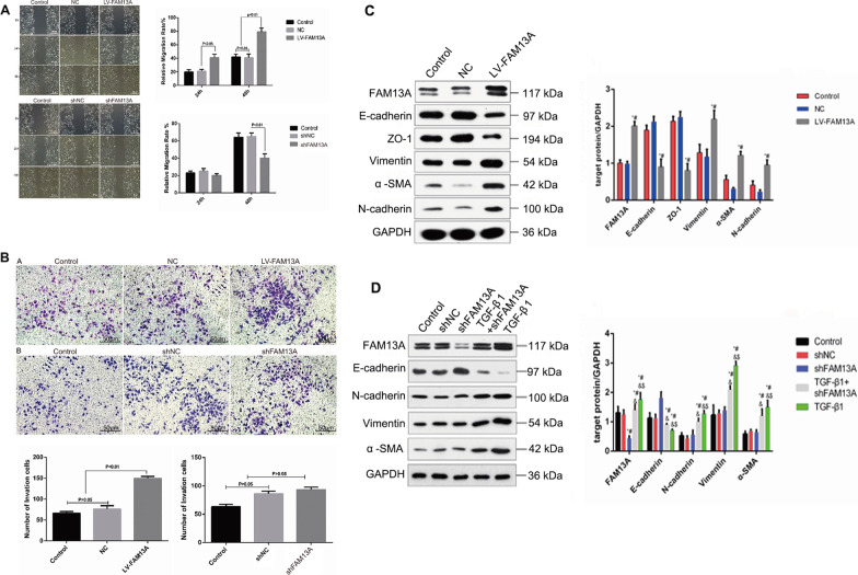
Results: Independent of smoking status, lung tissue samples from COPD patients exhibited significantly increased small airway thickness and collagen fiber deposition, along with enhanced protein levels of remodeling markers (collagen I, fibronectin, and MMP-9), mesenchymal markers (α-SMA, vimentin, and N-cadherin), TGF-β1, and FAM13A, compared with those from non-COPD patients. FAM13A expression negatively correlated with FEV1% and PO2 in COPD patients. In small airway epithelium, FAM13A expression negatively correlated with E-cadherin protein levels and positively correlated with vimentin protein levels. In BEAS-2B cells, TGF-β1 dose-dependently upregulated FAM13A protein levels. FAM13A overexpression significantly promoted cell proliferation and motility in BEAS-2B cells, whereas FAM13A silencing showed contrasting results. Furthermore, FAM13A knockdown partially reversed TGF-β1-induced EMT marker protein alterations in BEAS-2B cells.
Conclusions: FAM13A upregulation is associated with TGF-β1-induced EMT in the small airway epithelium of COPD patients independent of smoking status, serving as a potential therapeutic target for anti-EMT therapy in COPD.
Keywords: Chronic obstructive pulmonary disease; Epithelial–mesenchymal transition; FAM13A; Small airway epithelium; TGF-β.
Conflict of interest statement
The authors declare that they have no competing interests.
Figures
Similar articles
-
FAM13A as potential therapeutic target in modulating TGF-β-induced airway tissue remodeling in COPD.Am J Physiol Lung Cell Mol Physiol. 2021 Aug 1;321(2):L377-L391. doi: 10.1152/ajplung.00477.2020. Epub 2021 Jun 9. Am J Physiol Lung Cell Mol Physiol. 2021. PMID: 34105356
-
TWEAK enhances TGF-β-induced epithelial-mesenchymal transition in human bronchial epithelial cells.Respir Res. 2015 Apr 8;16(1):48. doi: 10.1186/s12931-015-0207-5. Respir Res. 2015. PMID: 25890309 Free PMC article.
-
Free radical generation induces epithelial-to-mesenchymal transition in lung epithelium via a TGF-β1-dependent mechanism.Free Radic Biol Med. 2012 Mar 15;52(6):1024-32. doi: 10.1016/j.freeradbiomed.2011.12.020. Epub 2012 Jan 2. Free Radic Biol Med. 2012. PMID: 22240154
-
The Role of Transforming Growth Factor-β (TGF-β) in Asthma and Chronic Obstructive Pulmonary Disease (COPD).Cells. 2024 Jul 29;13(15):1271. doi: 10.3390/cells13151271. Cells. 2024. PMID: 39120302 Free PMC article. Review.
-
Heparin-binding epidermal growth factor (HB-EGF) drives EMT in patients with COPD: implications for disease pathogenesis and novel therapies.Lab Invest. 2019 Feb;99(2):150-157. doi: 10.1038/s41374-018-0146-0. Epub 2018 Nov 19. Lab Invest. 2019. PMID: 30451982 Review.
Cited by
-
Systematic analysis of various RNA transcripts and construction of biological regulatory networks at the post-transcriptional level for chronic obstructive pulmonary disease.J Transl Med. 2023 Nov 7;21(1):790. doi: 10.1186/s12967-023-04674-7. J Transl Med. 2023. PMID: 37936118 Free PMC article.
-
The effects of epithelial-mesenchymal transitions in COPD induced by cigarette smoke: an update.Respir Res. 2022 Aug 31;23(1):225. doi: 10.1186/s12931-022-02153-z. Respir Res. 2022. PMID: 36045410 Free PMC article. Review.
-
FAM13A polymorphisms are associated with a specific susceptibility to clinical progression of oral cancer in alcohol drinkers.BMC Cancer. 2023 Jun 30;23(1):607. doi: 10.1186/s12885-023-11052-5. BMC Cancer. 2023. PMID: 37391706 Free PMC article.
-
The FAM13A Long Isoform Regulates Cilia Movement and Coordination in Airway Mucociliary Transport.Am J Respir Cell Mol Biol. 2024 Sep;71(3):282-293. doi: 10.1165/rcmb.2024-0063OC. Am J Respir Cell Mol Biol. 2024. PMID: 38691660 Free PMC article.
-
Genetic insights into idiopathic pulmonary fibrosis: a multi-omics approach to identify potential therapeutic targets.J Transl Med. 2025 Mar 16;23(1):337. doi: 10.1186/s12967-025-06368-8. J Transl Med. 2025. PMID: 40091050 Free PMC article.
References
-
- Lozano R, Naghavi M, Foreman K, Lim S, Shibuya K, Aboyans V, et al. Global and regional mortality from 235 causes of death for 20 age groups in 1990 and 2010: a systematic analysis for the Global Burden of Disease Study 2010. Lancet. 2012;380:2095–2128. doi: 10.1016/S0140-6736(12)61728-0. - DOI - PMC - PubMed
MeSH terms
Substances
Grants and funding
LinkOut - more resources
Full Text Sources
Other Literature Sources
Medical
Research Materials
Miscellaneous
