Review
. 2021 Sep:117:86-98.
doi: 10.1016/j.semcdb.2021.06.009. Epub 2021 Jun 29.

Spindle assembly checkpoint activation and silencing at kinetochores

Affiliations
Review

Spindle assembly checkpoint activation and silencing at kinetochores

Pablo Lara-Gonzalez et al. Semin Cell Dev Biol. 2021 Sep.

Abstract

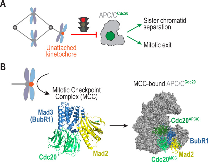
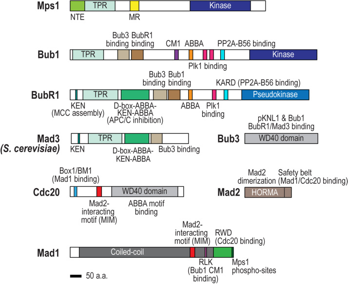
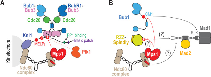
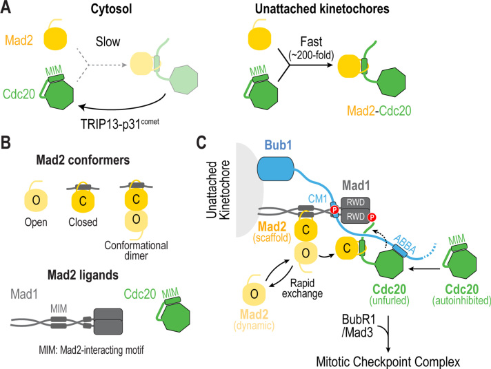
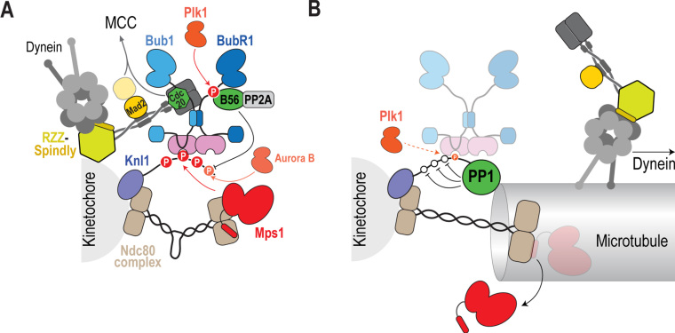
The spindle assembly checkpoint (SAC) is a surveillance mechanism that promotes accurate chromosome segregation in mitosis. The checkpoint senses the attachment state of kinetochores, the proteinaceous structures that assemble onto chromosomes in mitosis in order to mediate their interaction with spindle microtubules. When unattached, kinetochores generate a diffusible inhibitor that blocks the activity of the anaphase-promoting complex/cyclosome (APC/C), an E3 ubiquitin ligase required for sister chromatid separation and exit from mitosis. Work from the past decade has greatly illuminated our understanding of the mechanisms by which the diffusible inhibitor is assembled and how it inhibits the APC/C. However, less is understood about how SAC proteins are recruited to kinetochores in the absence of microtubule attachment, how the kinetochore catalyzes formation of the diffusible inhibitor, and how attachments silence the SAC at the kinetochore. Here, we summarize current understanding of the mechanisms that activate and silence the SAC at kinetochores and highlight open questions for future investigation.

Keywords: Aneuploidy; Catalysis; Checkpoint; Chromosome segregation; Kinetochores; Mitosis.


Figures

Fig. 1
Fig. 1
Schematic of Spindle Assembly Checkpoint (SAC) – dependent control of mitosis. (A) During mitosis, unattached kinetochores (orange) catalyze formation of an inhibitor that targets the APC/CCdc20, a ubiquitin ligase whose activity is essential for sister chromatid separation and mitotic exit. The SAC thereby ensures that the metaphase-anaphase transition occurs only after all kinetochores are attached to spindle microtubules. (B) The SAC functions by catalyzing the formation of the Mitotic Checkpoint Complex (MCC), which then binds APC/CCdc20 to block its activity. Shown here are the crystal structure of the MCC from S. pombe (PDB 4aez, left) and the cryo-EM structure of human APC/CCdc20 bound to the MCC (PDB 5lcw, right) . Note that Bub3, a component of the MCC in most species, is absent in these structures. (For interpretation of the references to color in this figure legend, the reader is referred to the web version of this article.)
Fig. 2
Fig. 2
Domain organization of SAC proteins. The SAC-relevant motifs/domains discussed in the text of Mps1, Bub1, Mad1, Mad2, BubR1/Mad3, Bub3, and Cdc20 are shown. Cartoons are based on the human proteins except for Mad3, which was based on the S. cerevisiae protein. Scale bar represents 50 amino acids.
Fig. 3
Fig. 3
Mechanisms for SAC protein recruitment at kinetochores. (A) The Bub1-Bub3-BubR1-Cdc20 module docks onto repetitive motifs in the Knl1 N-terminus that are phosphorylated by the kinase Mps1, which is recruited to kinetochores by the Ndc80 complex; Plk1 kinase activity targeting Knl1 also contributes to a varying extent, depending on the species. (B) Localization of the Mad1-Mad2 complex to unattached kinetochores is mediated by Bub1, the RZZ complex and the Ndc80-Mps1 module. Mps1 phosphorylates Bub1 CM1 to promote its interaction with Mad1 and may also target the RZZ complex; the Ndc80 complex may also contribute independently of recruiting Mps1. While not depicted here, the RZZ complex promotes expansion of the fibrous corona, the outermost region of the kinetochore, into crescents and rings that do not contain Bub1; thus, Bub1 and RZZ may recruit spatially distinct pools of Mad1-Mad2 complexes.
Fig. 4
Fig. 4
Catalysis of Mad2-Cdc20 complex formation at unattached kinetochores. (A) (left) Complex formation between Mad2 and Cdc20 is kinetically disfavored and subjected to disassembly by TRIP13-p31comet. (right) During mitosis, unattached kinetochores accelerate the rate of Mad2-Cdc20 complex formation by ~200 fold. (B) Mad2 has two different conformations, Open (O) and Closed (C), which interact with each other through dimerization. C-Mad2 is found bound to its ligands Mad1 and Mad2, which possess Mad2-interacting motifs (MIMs). (C) Proposed mechanism for how unattached kinetochores catalyze formation of the Mad2-Cdc20 complex. Mad2 is at two populations at the kinetochore: one that is stably bound to Mad1 (Mad2 scaffold) and a second that is cytosolic, transiently gets recruited to kinetochores through dimerization and becomes linked to Cdc20 (Mad2 dynamic). Kinetochores catalyze the transfer of Mad2 dynamic to Cdc20 by (1) recruiting Mad2 and Cdc20 to kinetochores, (2) positioning Cdc20 in close proximity to Mad2, and (3) unfurling Cdc20 to expose its MIM and promote the formation of the Mad2-Cdc20 complex.
Fig. 5
Fig. 5
SAC silencing at kinetochores. (A) At unattached kinetochores Mps1, anchored onto the Ndc80 complex, phosphorylates the Knl1 MELT repeats, which recruit the Bub module (Bub1, BubR1, Bub3 and Cdc20). Bub1, along with the RZZ complex, recruits the Mad1-Mad2 complex. The RZZ complex also recruits Spindly-Dynein. Phosphorylation of BubR1 by Plk1 promotes recruitment of the PP2A-B56 phosphatase, which opposes phosphorylation of the PP1 binding motif on Knl1 by Aurora B. (B) Microtubule attachments trigger the poleward transport of the Mad1-Mad2 complex by RZZ-Spindly-Dynein – this is a critical event in checkpoint silencing as preventing this transport leads to an active checkpoint despite kinetochore-microtubule attachment. In addition, microtubule attachment by Ndc80 causes either the displacement of Mps1 from kinetochores or intra-kinetochore stretching (depicted here for illustrative purposes as extension of the Ndc80 complex; we emphasize however that the molecular effector whose stretch affects SAC signaling is not known) which physically separates Mps1 from Knl1. Finally, PP1 recruitment causes dephosphorylation of the Knl1 MELTs to remove the Bub module from kinetochores. A small pool of the Bub module remains at attached kinetochores, which we speculate requires Plk1’s ability to also phosphorylate Knl1 MELTs.

References

    1. Musacchio A., Desai A. A molecular view of kinetochore assembly and function. Biology. 2017;6(1) - PMC - PubMed
    1. Lara-Gonzalez P., Westhorpe F.G., Taylor S.S. The spindle assembly checkpoint. Curr. Biol. 2012;22(22):R966–R980. - PubMed
    1. Jia L., Kim S., Yu H. Tracking spindle checkpoint signals from kinetochores to APC/C. Trends Biochem. Sci. 2013;38(6):302–311. - PubMed
    1. Musacchio A. The molecular biology of spindle assembly checkpoint signaling dynamics. Curr. Biol. 2015;25(20):R1002–R1018. - PubMed
    1. Corbett K.D. Molecular mechanisms of spindle assembly checkpoint activation and silencing. Prog. Mol. Subcell. Biol. 2017;56:429–455. - PubMed

Publication types