

Altered Blood Flow in the Ophthalmic and Internal Carotid Arteries in Patients with Age-Related Macular Degeneration Measured Using Noncontrast MR Angiography at 7T
- PMID: 34210664
- PMCID: PMC8423057
- DOI: 10.3174/ajnr.A7187
Altered Blood Flow in the Ophthalmic and Internal Carotid Arteries in Patients with Age-Related Macular Degeneration Measured Using Noncontrast MR Angiography at 7T
Abstract
Background and purpose: Age-related macular degeneration is associated with reduced perfusion of the eye; however, the role of altered blood flow in the upstream ophthalmic or internal carotid arteries is unclear. We used ultra-high-field MR imaging to investigate whether the diameter of and blood flow in the ophthalmic artery and/or the ICA are altered in age-related macular degeneration and whether any blood flow changes are associated with disease progression.
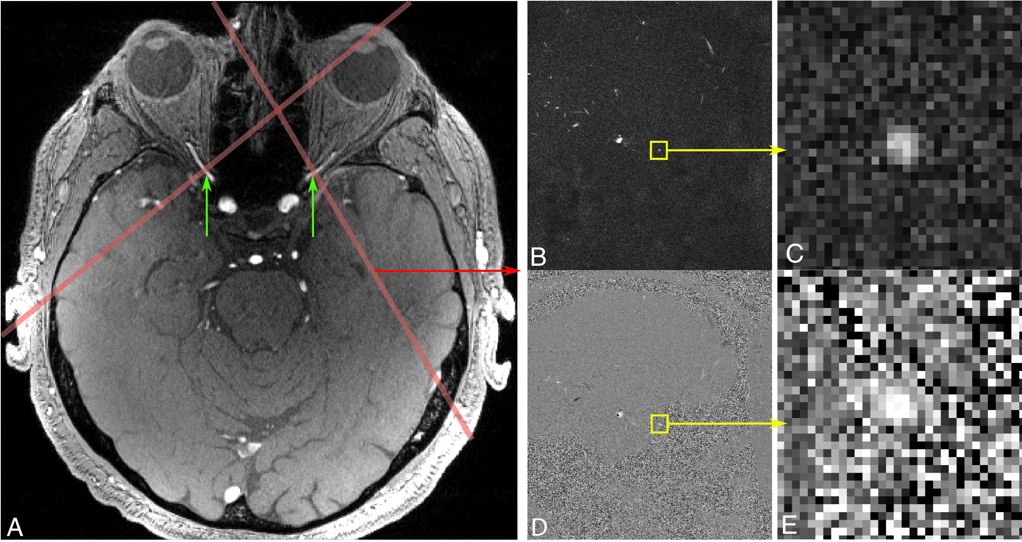
Materials and methods: Twenty-four patients with age-related macular degeneration and 13 similarly-aged healthy controls participated. TOF and high-resolution dynamic 2D phase-contrast MRA (0.26 × 0.26 × 2mm3, 100-ms effective sampling rate) was acquired at 7T. Vessel diameters were calculated from cross-sectional areas in phase-contrast acquisitions. Blood flow time-series were measured across the cardiac cycle.
Results: The ophthalmic artery vessel diameter was found to be significantly smaller in patients with age-related macular degeneration than in controls. Volumetric flow through the ophthalmic artery was significantly lower in patients with late age-related macular degeneration, with a significant trend of decreasing volumetric ophthalmic artery flow rates with increasing disease severity. The resistance index was significantly greater in patients with age-related macular degeneration than in controls in the ophthalmic artery. Flow velocity through the ophthalmic artery and ICA was significantly higher in patients with age-related macular degeneration. Ophthalmic artery blood flow as a percentage of ipsilateral ICA blood flow was nearly double in controls than in patients with age-related macular degeneration.
Conclusions: These findings support the hypothesis that vascular changes upstream to the eye are associated with the severity of age-related macular degeneration. Additional investigation into the potential causality of this relationship and whether treatments that improve ocular circulation slow disease progression is warranted.
© 2021 by American Journal of Neuroradiology.
Figures


Comment in
-
Regarding "Altered Blood Flow in the Ophthalmic and Internal Carotid Arteries in Patients with Age-Related Macular Degeneration Measured Using Noncontrast MR Angiography at 7T".AJNR Am J Neuroradiol. 2022 Dec;43(12):E60-E61. doi: 10.3174/ajnr.A7479. Epub 2022 Nov 24. AJNR Am J Neuroradiol. 2022. PMID: 36423953 No abstract available.
References
-
- Dry vs Wet Age-Related Macular Degeneration. AMDF. https://www.macular.org/dry-vs-wet-macular-degeneration. Accessed November 2, 2019
Publication types
MeSH terms
Grants and funding
LinkOut - more resources
Full Text Sources
Medical
Miscellaneous