. 2021 Aug 6;373(6555):648-654.
doi: 10.1126/science.abi7994. Epub 2021 Jul 1.

SARS-CoV-2 immune evasion by the B.1.427/B.1.429 variant of concern

Affiliations

SARS-CoV-2 immune evasion by the B.1.427/B.1.429 variant of concern

Matthew McCallum et al. Science. .

Abstract

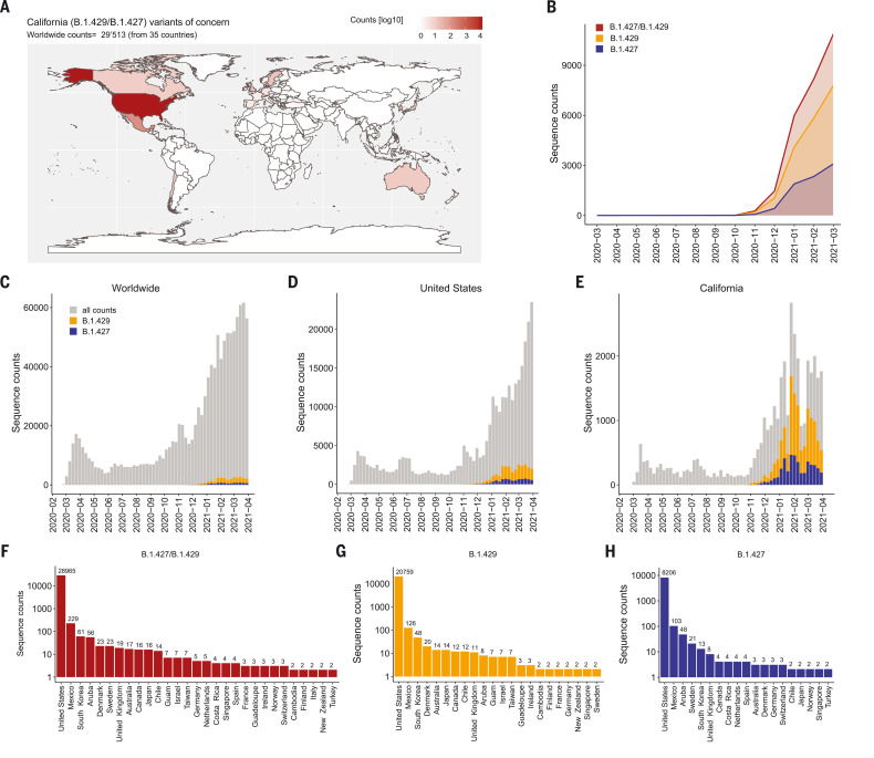
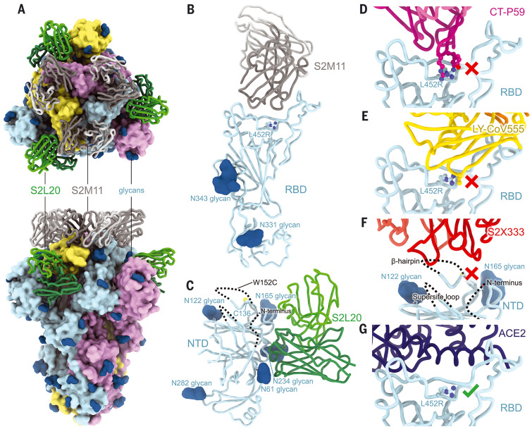
A novel variant of concern (VOC) named CAL.20C (B.1.427/B.1.429), which was originally detected in California, carries spike glycoprotein mutations S13I in the signal peptide, W152C in the N-terminal domain (NTD), and L452R in the receptor-binding domain (RBD). Plasma from individuals vaccinated with a Wuhan-1 isolate-based messenger RNA vaccine or from convalescent individuals exhibited neutralizing titers that were reduced 2- to 3.5-fold against the B.1.427/B.1.429 variant relative to wild-type pseudoviruses. The L452R mutation reduced neutralizing activity in 14 of 34 RBD-specific monoclonal antibodies (mAbs). The S13I and W152C mutations resulted in total loss of neutralization for 10 of 10 NTD-specific mAbs because the NTD antigenic supersite was remodeled by a shift of the signal peptide cleavage site and the formation of a new disulfide bond, as revealed by mass spectrometry and structural studies.


Figures

Fig. 1.
Fig. 1.. Geographic distribution and evolution of incidence over time of the SARS-CoV-2 B.1.427/B.1.429 VOC.
(A) World map showing the geographic distribution and sequence counts of B.1.427/B.1.429 VOC as of 30 April 2021. (B) Cumulative and individual B.1.427/B.1.429 VOC sequence counts by month. (C to E) Total number of SARS-CoV-2 (gray) and B.1.427/B.1.429 VOC (blue/orange) sequences deposited on a monthly basis worldwide (C), in the United States (D), and in California (E). (F to H) Total number of B.1.427/B.1.429 (F), B.1.429 (G), and B.1.427 (H) sequences deposited by country as of 30 April 2021. Only countries with two or more deposited sequences are shown.
Fig. 2.
Fig. 2.. B.1.427/B.1.429 S pseudotyped virus neutralization by vaccine-elicited and COVID-19 convalescent plasma.
(A, B, E, and F) Neutralizing Ab titers [mean inhibition dilution (ID50)] shown as pairwise connected [(A) and (E)] or GMT [(B) and (F)] against MLV [(A) and (B)] or VSV [(E) and (F)] pseudotyped viruses harboring G614 SARS-CoV-2 S or B.1.427/B.1.429 (B.1.429) S determined using plasma from individuals who received two doses of the Moderna mRNA-1273 vaccine (blue). (C, D, G, and H) Neutralizing Ab titers (ID50) shown as pairwise connected [(C) and (G)] or GMT [(D) and (H)] against MLV [(C) and (D)] or VSV [(G) and (H)] pseudotyped viruses harboring G614 SARS-CoV-2 S or B.1.427/B.1.429 (B.1.429) S determined using plasma from individuals who received two doses of the Pfizer/BioNtech BNT162b2 mRNA vaccine (red). (I and J) Neutralizing Ab ID50 (I) and GMT (J) titers against VSV pseudotyped viruses harboring D614 SARS-CoV-2 S, B.1.427/B.1.429 S, B.1.1.7 S, B.1.351 S, or P.1 S determined using plasma from naïve (blue) and previously infected (red) individuals who received two doses of the Pfizer/BioNtech BNT162b2 mRNA vaccine. “Naïve” indicates vaccinated individuals who had not been previously infected with SARS-CoV-2; “immune” refers to vaccinated individuals who had been previously infected with SARS-CoV-2. (K and L) Neutralizing Ab ID50 (K) and GMT (L) titers against VSV pseudotyped viruses harboring D614 SARS-CoV-2 S, B.1.427/B.1.429 S, B.1.1.7 S, B.1.351 S, or P.1 S determined using plasma from convalescent individuals who were infected with WT SARS-CoV-2. Neutralization data shown in (A) to (H) and (I) to (L) were performed using 293T-ACE2 and VeroE6-TMPRSS2, respectively. Data are average of n = 2 replicates.
Fig. 3.
Fig. 3.. Neutralization by a panel of RBD- and NTD-specific mAbs against SARS-CoV-2 D614 S and B.1.427/B.1.429 S pseudoviruses.
(A and D) Neutralization of SARS-CoV-2 pseudotyped VSV carrying D614 (gray) or B.1.427/B.1.429 (orange) S protein by clinical stage RBD mAbs (A) and an NTD-targeting mAb (S2X333) (D). Data are representative of n = 2 replicates. (B and E) Neutralization of SARS-CoV-2 S VSV pseudotypes carrying D614 or B.1.427/B.1.429 S by 34 mAbs targeting the RBD (B) and 10 mAbs targeting the NTD (E). Data are the mean of 50% inhibitory concentration (IC50) values (in nanograms per milliliter) of n = 2 independent experiments. Non-neutralizing IC50 titers were set at 105 ng/ml. (C and F) Neutralization by RBD-specific (C) and NTD-specific (F) mAbs shown as mean IC50 values (top) and mean fold change (bottom) for B.1.427/B.1.429 S (orange) relative to D614 S (gray) VSV pseudoviruses. VIR-7831 is a derivative of S309 mAb (sotrovimab). *VIR-7832 (variant of VIR-7831 carrying the LS-GAALIE Fc mutations) shown as squares. Non-neutralizing IC50 titers and fold change were set to 105 ng/ml and 104, respectively.
Fig. 4.
Fig. 4.. CryoEM structure of the SARS-CoV-2 B.1.427/B.1.429 S ectodomain trimer.
(A) Structure of the S trimer (surface rendering) bound to the S2M11 and S2L20 Fabs (ribbons) in two orthogonal orientations. SARS-CoV-2 S protomers are colored pink, cyan, and gold, and the S2L20 Fab heavy and light chains are colored dark and light green, respectively, and the S2M11 Fab heavy and light chains are colored dark and light gray, respectively. Only the Fab-variable domains are resolved in the map. N-linked glycans are rendered as dark blue spheres. (B) Magnified view of the S2M11-bound RBD with R452 shown in ball and stick representation. (C) Magnified view of the S2L20-bound NTD with disordered N terminus, supersite β-hairpin, and loop regions shown as dashed lines. (D) Superimposition of the CT-P59–bound SARS-CoV-2 RBD structure (PDB 7CM4) on the SARS-CoV-2 B.1.427/B.1.429 S cryoEM structure showing that R452 would sterically clash with the mAb. (E) Superimposition of the LY-CoV555–bound SARS-CoV-2 RBD structure (PDB 7KMG) on the SARS-CoV-2 B.1.427/B.1.429 S cryoEM structure showing that L452R would sterically clash with the mAb. (F) Superimposition of the S2X333-bound SARS-CoV-2 S structure (PDB 7LXW) on the SARS-CoV-2 B.1.427/B.1.429 S cryoEM structure showing that most of the NTD antigenic supersite epitope residues are disordered. (G) Superimposition of the ACE2-bound SARS-CoV-2 RBD structure (PDB 7DMU) on the SARS-CoV-2 B.1.427/B.1.429 S cryoEM structure showing that L452R points away from the interface with ACE2.
Fig. 5.
Fig. 5.. The B.1.427/B.1.429 S S13I and W152C mutations lead to immune evasion.
(A) Binding of a panel of 11 neutralizing (antigenic site i) and one non-neutralizing (antigenic site iv) NTD-specific mAbs to recombinant SARS-CoV-2 NTD variants analyzed by ELISA displayed as a heatmap. (B) Binding of plasma Abs from vaccinated individuals to recombinant SARS-CoV-2 NTD variants analyzed by ELISA. The mean dilution factor for each mutant was compared by the one-way ANOVA test against WT (*P < 0.05, **P < 0.001). (C to G) Deconvoluted mass spectra of purified NTD constructs, including the WT NTD with the native signal peptide (C), the S13I NTD (D), the S13I and W152C NTD (E), the W152C NTD (F), and the S12F NTD (G). The empirical mass (black) and theoretical mass (red) are shown beside the corresponding peak. An additional 119 Da were observed for the S13I and W152C NTDs, corresponding to cysteinylation of the free cysteine residue in these constructs (as L-cysteine was present in the expression media). The cleaved signal peptide (blue text) and subsequent residue sequence (black text) are also shown based on the MS results. Mutated residues are shown in bold. Cysteines are highlighted in light orange (unless in the cleaved signal peptide), and disulfide bonds are shown as dotted light orange lines between cysteines. Residues are numbered for reference.

Update of

  • SARS-CoV-2 immune evasion by variant B.1.427/B.1.429.
    McCallum M, Bassi J, Marco A, Chen A, Walls AC, Iulio JD, Tortorici MA, Navarro MJ, Silacci-Fregni C, Saliba C, Agostini M, Pinto D, Culap K, Bianchi S, Jaconi S, Cameroni E, Bowen JE, Tilles SW, Pizzuto MS, Guastalla SB, Bona G, Pellanda AF, Garzoni C, Van Voorhis WC, Rosen LE, Snell G, Telenti A, Virgin HW, Piccoli L, Corti D, Veesler D. McCallum M, et al. bioRxiv [Preprint]. 2021 Apr 1:2021.03.31.437925. doi: 10.1101/2021.03.31.437925. bioRxiv. 2021. Update in: Science. 2021 Aug 6;373(6555):648-654. doi: 10.1126/science.abi7994. PMID: 33821281 Free PMC article. Updated. Preprint.

References

    1. Walls A. C., Park Y. J., Tortorici M. A., Wall A., McGuire A. T., Veesler D., Structure, function, and antigenicity of the SARS-CoV-2 spike glycoprotein. Cell 181, 281–292.e6 (2020). 10.1016/j.cell.2020.02.058 - DOI - PMC - PubMed
    1. Wrapp D., Wang N., Corbett K. S., Goldsmith J. A., Hsieh C. L., Abiona O., Graham B. S., McLellan J. S., Cryo-EM structure of the 2019-nCoV spike in the prefusion conformation. Science 367, 1260–1263 (2020). 10.1126/science.abb2507 - DOI - PMC - PubMed
    1. Walls A. C., Tortorici M. A., Bosch B. J., Frenz B., Rottier P. J. M., DiMaio F., Rey F. A., Veesler D., Cryo-electron microscopy structure of a coronavirus spike glycoprotein trimer. Nature 531, 114–117 (2016). 10.1038/nature16988 - DOI - PMC - PubMed
    1. Tortorici M. A., Veesler D., Structural insights into coronavirus entry. Adv. Virus Res. 105, 93–116 (2019). 10.1016/bs.aivir.2019.08.002 - DOI - PMC - PubMed
    1. Letko M., Marzi A., Munster V., Functional assessment of cell entry and receptor usage for SARS-CoV-2 and other lineage B betacoronaviruses. Nat. Microbiol. 5, 562–569 (2020). 10.1038/s41564-020-0688-y - DOI - PMC - PubMed

Publication types

MeSH terms