. 2021 Jul 1;11(1):13705.
doi: 10.1038/s41598-021-91662-w.

Concurrent mutations in RNA-dependent RNA polymerase and spike protein emerged as the epidemiologically most successful SARS-CoV-2 variant

Affiliations

Concurrent mutations in RNA-dependent RNA polymerase and spike protein emerged as the epidemiologically most successful SARS-CoV-2 variant

Sten Ilmjärv et al. Sci Rep. .

Abstract

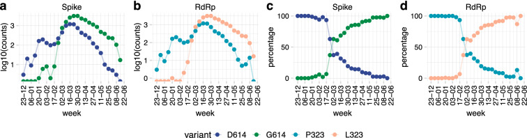
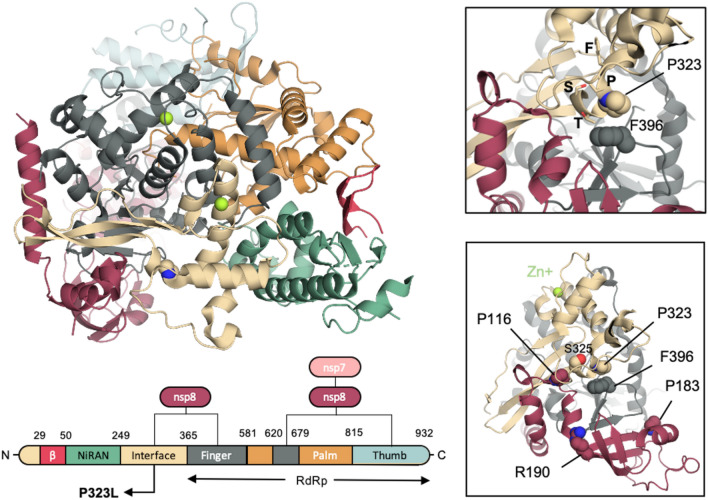
The D614G mutation in the Spike protein of the SARS-CoV-2 has effectively replaced the early pandemic-causing variant. Using pseudotyped lentivectors, we confirmed that the aspartate replacement by glycine in position 614 is markedly more infectious. Molecular modelling suggests that the G614 mutation facilitates transition towards an open state of the Spike protein. To explain the epidemiological success of D614G, we analysed the evolution of 27,086 high-quality SARS-CoV-2 genome sequences from GISAID. We observed striking coevolution of D614G with the P323L mutation in the viral polymerase. Importantly, the exclusive presence of G614 or L323 did not become epidemiologically relevant. In contrast, the combination of the two mutations gave rise to a viral G/L variant that has all but replaced the initial D/P variant. Our results suggest that the P323L mutation, located in the interface domain of the RNA-dependent RNA polymerase, is a necessary alteration that led to the epidemiological success of the present variant of SARS-CoV-2. However, we did not observe a significant correlation between reported COVID-19 mortality in different countries and the prevalence of the Wuhan versus G/L variant. Nevertheless, when comparing the speed of emergence and the ultimate predominance in individual countries, it is clear that the G/L variant displays major epidemiological supremacy over the original variant.


Conflict of interest statement

MC worked for Neurix SA. All authors declare no competing interests.

Figures

Figure 1
Figure 1
Structural implications of the D614G mutation in the S-protein. (a) Top and side views of the S-protein in the closed and open conformations. D614 in the three protomers is depicted in orange van der Waals spheres. The RBD of protomer A in the down and up state is coloured in light green and dark pink, respectively. Graphical representation of the domain organisation of the S-protein: signal peptide (SP), receptor binding domain (RBD), N-terminal domain (NTD), subdomains 1 and 2 (SD1 and SD2), fusion peptide (FP), heptad repeat 1 (HR1), central helix (CH), connector domain (CD), heptad repeat 2 (HR2), transmembrane domain (TM). (b) Time series of D614 with K835 and P826 at the A–C interface over the course of the simulations initiated from the closed (green) and partially open (pink) conformations. The equivalent interactions of D614 at the B–C interface are given in grey. The probability distribution function (PDF) of the two interactions are displayed on the top of each timeseries. (c) Upper panels: interactions involving D614 for the closed (left) and open (right) conformation at the end of each simulation. The D614 residue of chain A is depicted in pink and green spheres and the interacting residues of chain C are represented in white spheres. Bottom-panels: interactions involving D614, when mutated to glycine, for the closed (left) and open (right) conformation at the end of each simulation. The Cα atom of G614 of chain A is depicted in pink and green spheres and the interacting residues of chain C are represented in white spheres.
Figure 2
Figure 2
SARS-CoV-2 S-protein D614G variant shows increased infectivity. (a,b) Real-time RT-PCR for ACE2, TMPRSS2, furin and cathepsin L mRNA expression profiles in HeLa (a) and A549 (b) cells. (cf) Spike pseudotyped lentiviral infectivity assays. Pseudotyped lentiviruses expressing Luciferase and GFP and pseudotyped with SARS-CoV-2-D614 or G614 full length or truncated version were used to transduce HeLa and A549 overexpressing or not either ACE2 and/or TMPRSS2. (c,d) Luciferase assay of HeLa (c) and A549 (d) at 5 days post transduction with 80 μL of SARS-CoV-2 spike pseudotyped lentivirus. (e,f) Percent of GFP + assay of HeLa (e) and A549 (f) cells at 5 days post transduction with 80 μL of SARS-CoV-2 S-protein pseudotyped lentivirus analysed by Flow cytometry. The experiments were done in triplicates and repeated three times with three independent lentiviral stocks that was resulting from independent transfection. One representative is shown with error bars indicating SEM of replicates.
Figure 3
Figure 3
Two major protein variants of SARS-CoV-2: S protein D614G and RNA-dependent polymerase P323L. Available high-quality sequences of SARS-CoV-2 (27,084) were analysed for the D614 and G614 variants of the S-protein and the P323 and L323 of the RdR polymerase. Data are shown as total number of sequences per week (a, note the logarithmic scale) or percent of sequences per week (b).
Figure 4
Figure 4
Temporal and spatial distribution of the four possible D614G and P323L combinations. Available high-quality sequences of SARS-CoV-2 (27,075) were analysed. (a) G614/L323 (double mutant) represented more than two-thirds, and D614/P323 (the original Wuhan sequence) about one-third of sequences; the single mutant variants G614/P323 and D614/L323 were very rare, (b) weekly number of variants over time; (c) cumulative number of variants over time. (d) Percentage of variants in different countries; only countries with at least 20 sequences are represented.
Figure 5
Figure 5
Temporal evolution of the four possible D614G and P323L variants in different countries. Available high-quality sequences of SARS-CoV-2 (27 075) were analysed for the different possible combinations of the D614 and G614 variants of the spike protein and the P323 and L323 of the RdR polymerase based on country of origin of the sequences. Only countries with at least 20 sequences were included in this analysis. Results are shown as cumulative number of sequences over time (the weekly number of sequences is provided as a Supplementary Figure 4).
Figure 6
Figure 6
SARS-CoV-2 variants: mortality, case fatality rate, and patient status. Relationship between the percentage of the two main SARS-CoV-2 variants (D614/P323 and G614/L323) in different countries and (a) number of deaths per 100 000 population and (b) case fatality rate in the respective countries. Numbers of deaths per 100 000 population and case fatality rates were taken from the Johns Hopkins Coronavirus Resource Center (coronavirus.jhu.edu/data/mortality) at 4th of August 2020.
Figure 7
Figure 7
Comparative expansion of SARS-CoV-2 variants in different countries: (a) time of first reported D614/P323 sequence versus first reported G614/L323 sequence (blue bars: D614/P323 appeared before G614/L323; black bars: D614/P323 appeared before G614/L323). (b) Percent of sequences of a given variant in the 14-day time period after its first appearance in a given country (100% is total number of sequences during the 14-day time period). Only countries with at least 5 sequences collected during 14 days after the appearance of first sequence are used (c) mean ± SEM of emergence of the variants in different countries with at least 5 sequences within the first 14 days.
Figure 8
Figure 8
Structural implications of the D614G mutation in S-protein. (a) Domain organisation of SARS-CoV-2 nsp12 in complex with nsp7 and nsp8 cofactors. The P323 is depicted in beige spheres. (b) Close up around P323. The residues of the 321FPPTS325 stretch are depicted in sticks, while F396 that P323 interacts with is depicted in spheres. (c) Residues P116, P183 and R190 of the nsp8 that has been reported to be crucial for the nsp8/nsp12 interaction in SAR-CoV-1 are depicted in red spheres.

References

    1. Petersen E, et al. Comparing SARS-CoV-2 with SARS-CoV and influenza pandemics. Lancet. Infect. Dis. 2020 doi: 10.1016/s1473-3099(20)30484-9. - DOI - PMC - PubMed
    1. Smith EC, Blanc H, Surdel MC, Vignuzzi M, Denison MR. Coronaviruses lacking exoribonuclease activity are susceptible to lethal mutagenesis: evidence for proofreading and potential therapeutics. PLoS Pathog. 2013;9:e1003565. doi: 10.1371/journal.ppat.1003565. - DOI - PMC - PubMed
    1. Ferron F, et al. Structural and molecular basis of mismatch correction and ribavirin excision from coronavirus RNA. Proc. Natl. Acad. Sci. USA. 2018;115:E162–E171. doi: 10.1073/pnas.1718806115. - DOI - PMC - PubMed
    1. Denison MR, Graham RL, Donaldson EF, Eckerle LD, Baric RS. Coronaviruses: an RNA proofreading machine regulates replication fidelity and diversity. RNA Biol. 2011;8:270–279. doi: 10.4161/rna.8.2.15013. - DOI - PMC - PubMed
    1. Bar-On YM, Flamholz A, Phillips R, Milo R. SARS-CoV-2 (COVID-19) by the numbers. Elife. 2020 doi: 10.7554/elife.57309. - DOI - PMC - PubMed

Publication types

MeSH terms

Substances