. 2021 Jul 1;11(1):13700.
doi: 10.1038/s41598-021-93156-1.

Canagliflozin protects against sepsis capillary leak syndrome by activating endothelial α1AMPK

Affiliations

Canagliflozin protects against sepsis capillary leak syndrome by activating endothelial α1AMPK

Marine Angé et al. Sci Rep. .

Abstract

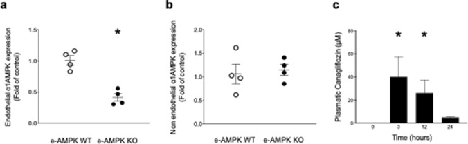
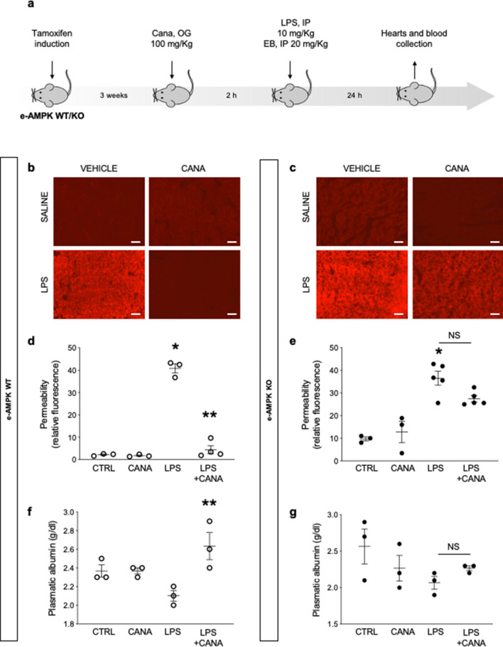
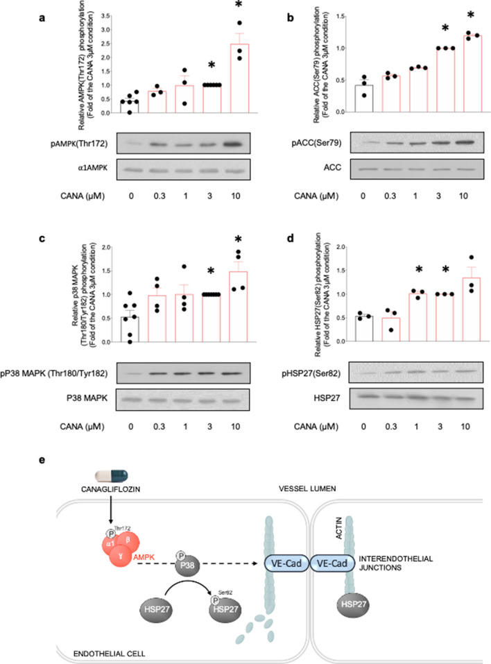
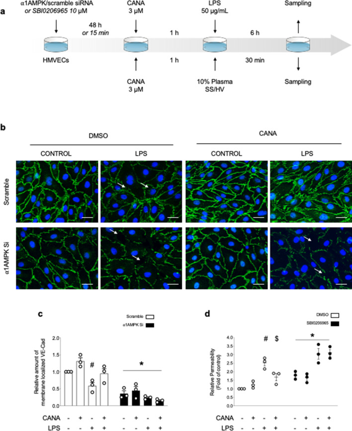
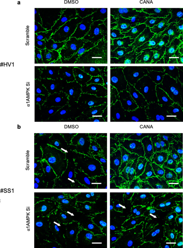
Sepsis capillary leak syndrome (SCLS) is an independent prognostic factor for poor sepsis outcome. We previously demonstrated that α1AMP-activated protein kinase (α1AMPK) prevents sepsis-induced vascular hyperpermeability by mechanisms involving VE-cadherin (VE-Cad) stabilization and activation of p38 mitogen activated protein kinase/heat shock protein of 27 kDa (p38MAPK/HSP27) pathway. Canagliflozin, a sodium-glucose co-transporter 2 inhibitor, has recently been proven to activate AMPK in endothelial cells. Therefore, we hypothesized that canagliflozin could be of therapeutic potential in patients suffering from SCLS. We herein report that canagliflozin, used at clinically relevant concentrations, counteracts lipopolysaccharide-induced vascular hyperpermeability and albumin leakage in wild-type, but not in endothelial-specific α1AMPK-knockout mice. In vitro, canagliflozin was demonstrated to activate α1AMPK/p38MAPK/HSP27 pathway and to preserve VE-Cad's integrity in human endothelial cells exposed to human septic plasma. In conclusion, our data demonstrate that canagliflozin protects against SCLS via an α1AMPK-dependent pathway, and lead us to consider novel therapeutic perspectives for this drug in SCLS.


Conflict of interest statement

The authors declare no competing interests.

Figures

Figure 1
Figure 1
Generation and validation of the experimental model. e-AMPK WT/KO mice were intraperitoneally administered Tamoxifen (500 µg/mice) for 5 consecutive days in order to induce ⍺1AMPK invalidation specifically in the endothelium. 3 weeks after the last Tamoxifen injection, mice were sacrificed and endothelial cells were immunoprecipitated from lung tissue with dynabeads. ⍺1AMPK gene expression was detected by TaqMan qPCR on both isolated endothelial cells and supernatants. (a) Validation of endothelial ⍺1AMPK deletion’s extent in e-AMPK KO. ⍺1AMPK expression detected by TaqMan qPCR in immunoprecipitated endothelial cells. (b) Validation of endothelial ⍺1AMPK deletion’s specificity. ⍺1AMPK expression detected by TaqMan qPCR on lysates of lung tissue depleted of endothelial cells. (c) Time course of canagliflozin plasma levels in mice treated by oral gavage, 100 mg/kg, during indicated time. The data are mean ± SEM, n = 3 to 5/group. The data were analyzed using one-way ANOVA test.
Figure 2
Figure 2
Canagliflozin protects against lipopolysaccharide induced capillary leak syndrome via endothelial ⍺1AMPK dependent mechanisms. (a) Schematic representation of the experimental protocol for in vivo permeability assessment. e-AMPK WT/KO mice were intraperitoneally administered Tamoxifen (500 µg/mice) for five consecutive days in order to induce ⍺1AMPK invalidation specifically in the endothelium. 3 weeks after the last Tamoxifen injection, mice were treated with canagliflozin (Cana) (100 mg/Kg) by oral gavage, two hours before being submitted to lipopolysaccharide (LPS) treatment (sublethal doses, 10 mg/kg) by intraperitoneal (IP) injection. Evans Blue Dye (EBD) was administered by IP simultaneously with LPS. (be) Cardiac vascular permeability was assessed via EBD fluorescence quantification on myocardial sections of hearts sampled 24 h after injection of LPS or saline vehicle. Representative images (b, c) and quantifications (d, e) are shown. Scale bar, 200 µm. (f, g) Plasmatic albumin levels were measured on blood samples collected 24 h after injection of LPS or saline vehicle. The data are mean ± SEM, n = 3 to 5/group. *p < 0.05 is relative to control saline group, **p < 0.05 is relative to LPS treated group. NS = nonsignificant. The data underwent two-way ANOVA.
Figure 3
Figure 3
Canagliflozin activates the ⍺1AMPK/p38MAPK/HSP27 pathway in HMECs. HMECs were treated with canagliflozin (Cana) for the indicated concentrations during one hour. Cell lysates were submitted to western blot analysis and probed with total and phosphorylated (a) ⍺1AMPK (Thr172), (b) ACC (Ser79), (c) p38 MAPK (Thr180/Tyr182) and (d) HSP27(Ser82) antibodies, (e) Molecular mechanisms underlying the protective action of canagliflozin on interendothelial junctions. Representative western blots and quantification are shown. Data are fold of the 3 µM condition and expressed as mean ± SEM (3 to 6 biological replicates for each condition). *p < 0.05 is relative to untreated HMECs. The data underwent one-way ANOVA.
Figure 4
Figure 4
AMPK activation by Canagliflozin protects VE-Cad organization and endothelial barrier function against LPS injury. (a) Schematic representation of the experimental protocol. For (b, c), HMECs were transfected with scramble or ⍺1AMPK targeting siRNA (50 nM) for 48 h, then treated with canagliflozin (Cana) (3 µM) or DMSO for one hour, before adding lipopolysaccharide (LPS) or vehicle (50 µg/mL) for 6 h. For (d), SBI compound was incubated for 15 min, then canagliflozin (3 µM) or DMSO were incubated for one hour, before adding LPS (50 µg/mL) for 6 h. (b, c) VE-Cad immunostainings were performed on HMECs treated according to the protocol detailed in (a). Representative images (b) and quantifications (c) are shown. Intercellular gaps are indicated by white arrows. Nuclei were stained with DAPI. Scale bar, 50 µm. (d) Endothelial permeability in response to canagliflozin, LPS challenge, and AMPK inhibitor SBI0206965. HMECs were grown on gelatin-coated Transwell inserts for 72 h and treated according to the protocol detailed in (a). Data are expressed as mean ± SEM (3 biological replicates for each condition). #p < 0.05 is relative to respective non-treated HMECs, $p < 0.05 is relative to LPS-only treated HMECs, and *p < 0.05 is relative to cells treated with DMSO. The data underwent two-way ANOVA.
Figure 5
Figure 5
AMPK activation by Canagliflozin protects VE-Cad integrity in HMECs challenged with human septic plasma. VE-Cad immunostainings performed on HMECs. HMECs were transfected with scramble or ⍺1AMPK targeting siRNA (50 nM) for 48 h, before being treated with canagliflozin (3 µM) or DMSO for one hour, then incubated with 10% plasma of (a) healthy volunteers (HV) or (b) septic shock patients (SS) for 30 min. Typical examples of pictures are shown. The experiment has been repeated with 4 different donors for each group (see Supplemental Fig. 2). Intercellular gaps are indicated by white arrows. Nuclei were stained with DAPI. Scale bar, 50 µm.

References

    1. Rudd KE, et al. Global, regional, and national sepsis incidence and mortality, 1990–2017: Analysis for the Global Burden of Disease Study. Lancet. 2020;395:200–211. doi: 10.1016/s0140-6736(19)32989-7. - DOI - PMC - PubMed
    1. Singer M, et al. The third international consensus definitions for sepsis and septic shock (Sepsis-3) JAMA. 2016;315:801–810. doi: 10.1001/jama.2016.0287. - DOI - PMC - PubMed
    1. Fleischmann C, et al. Assessment of global incidence and mortality of hospital-treated sepsis. Current estimates and limitations. Am. J. Respir. Crit. Care Med. 2016;193:259–272. doi: 10.1164/rccm.201504-0781OC. - DOI - PubMed
    1. Acheampong A, Vincent JL. A positive fluid balance is an independent prognostic factor in patients with sepsis. Crit. Care. 2015;19:251. doi: 10.1186/s13054-015-0970-1. - DOI - PMC - PubMed
    1. Castanares-Zapatero D, et al. Connection between cardiac vascular permeability, myocardial edema, and inflammation during sepsis: role of the alpha1AMP-activated protein kinase isoform. Crit. Care Med. 2013;41:e411–422. doi: 10.1097/CCM.0b013e31829866dc. - DOI - PubMed

Publication types

MeSH terms

Substances