. 2021 Jul 1;11(1):13674.
doi: 10.1038/s41598-021-93256-y.

Local application of Usag-1 siRNA can promote tooth regeneration in Runx2-deficient mice

Affiliations

Local application of Usag-1 siRNA can promote tooth regeneration in Runx2-deficient mice

Sayaka Mishima et al. Sci Rep. .

Abstract

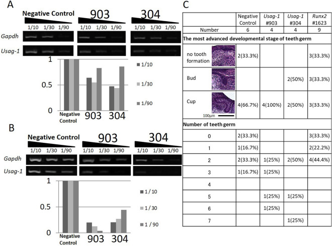
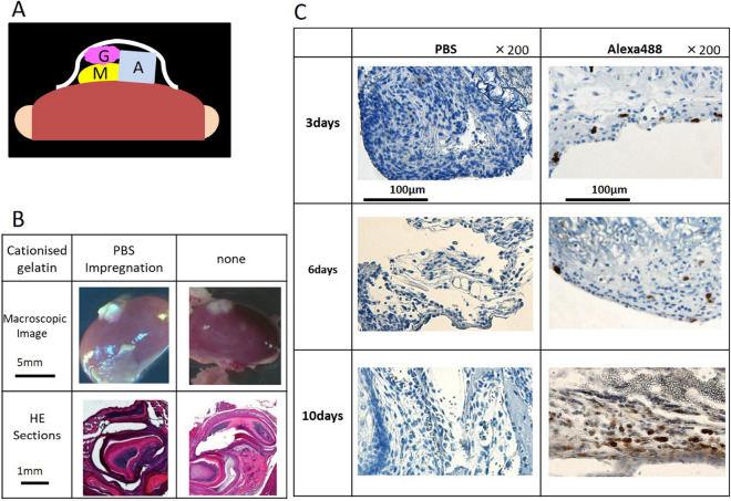
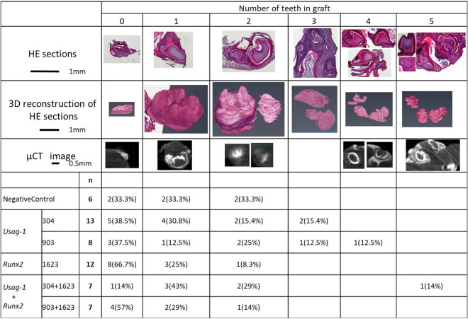
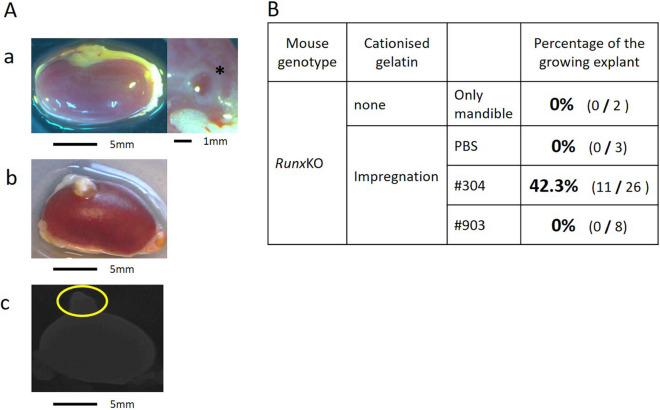
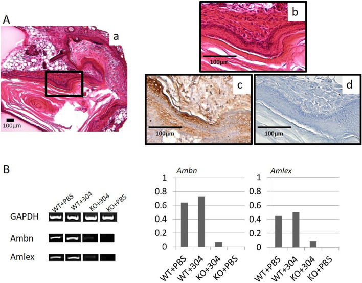
Runt-related transcription factor 2 (Runx2)-deficient mice can be used to model congenital tooth agenesis in humans. Conversely, uterine sensitization-associated gene-1 (Usag-1)-deficient mice exhibit supernumerary tooth formation. Arrested tooth formation can be restored by crossing both knockout-mouse strains; however, it remains unclear whether topical inhibition of Usag-1 expression can enable the recovery of tooth formation in Runx2-deficient mice. Here, we tested whether inhibiting the topical expression of Usag-1 can reverse arrested tooth formation after Runx2 abrogation. The results showed that local application of Usag-1 Stealth small interfering RNA (siRNA) promoted tooth development following Runx2 siRNA-induced agenesis. Additionally, renal capsule transplantation of siRNA-loaded cationized, gelatin-treated mouse mandibles confirmed that cationized gelatin can serve as an effective drug-delivery system. We then performed renal capsule transplantation of wild-type and Runx2-knockout (KO) mouse mandibles, treated with Usag-1 siRNA, revealing that hindered tooth formation was rescued by Usag-1 knockdown. Furthermore, topically applied Usag-1 siRNA partially rescued arrested tooth development in Runx2-KO mice, demonstrating its potential for regenerating teeth in Runx2-deficient mice. Our findings have implications for developing topical treatments for congenital tooth agenesis.


Conflict of interest statement

The authors declare no competing interests.

Figures

Figure 1
Figure 1
Silencing with Usag-1 and Runx2 Stealth siRNAs and the effects on tooth formation. (A) Usag-1 expression in mHAT9d cells at 48 h post-transfection of Usag-1 Stealth siRNAs (#903 and #304), as determined by sqRT-PCR. Both siRNAs showed ~ 50% knockdown efficiency. The synthesized complementary DNA (cDNA) samples were serially diluted and subjected to semi-quantitative PCR analysis. 1/10, 1/30, and 1/90 indicates dilution ratio. (B) Usag-1 expression in organ cultures treated with Usag-1 Stealth siRNAs (#903 and #304), as determined by sqRT-PCR. The knockdown efficiencies of both siRNAs in E10 mandible explant cultures were comparable to those in mHAT9d cells. (C) The most advanced developmental stage and number of tooth germs in organ cultures 10 days after co-administration of Usag-1 and Runx2 Stealth siRNAs. Developmental stages were classified as no tooth formation, the bud stage, or the cup stage.
Figure 2
Figure 2
Renal capsule assays using lateral mandibles from wild-type mice. (A) Schematic illustration of the procedure used for renal capsule transplantation. M indicates the lateral mandibles of E10 mice, G indicates cationized gelatin containing Stealth siRNA, and A indicates agarose agar (was used to maintain the graft space). (B) We evaluated H&E-stained sections, where the lateral mandible from wild-type mice was transplanted beneath the kidney capsule of nude mice (KSN/Slc). Renal capsule assays were performed with two groups: lateral mandibles transplanted with or without a cationized gelatin sheet containing PBS. Based on histochemical staining, we observed that the sections contained tooth structures, although we found no differences in the results with or without cationized gelatin. (C) Histological sections at days 3, 6, and 10 post -transplantation. Sections were evaluated by immunostaining. Magnification, 200× . Cells were immunostained with an Alexa Fluor 488-conjugated antibody.
Figure 3
Figure 3
The number of teeth in a graft at 19 days post -transplantation. We performed renal capsule transplantations of wild-type mouse mandibles together with cationized gelatin sheets containing (1) a negative-control siRNA, (2) Usag-1 siRNA or Runx2 siRNA, or (3) Usag-1 siRNA + Runx2 siRNA. The number of tooth structures was determined by histological evaluation of serial H&E sections and 3D micro-CT analysis. The number of tooth structures was confirmed by 3D reconstruction of serial H&E sections in representative samples. P = 0.020 for Usag-1 #304 vs. Runx2 Stealth siRNA; P = 0.656 for Usag-1 #903 vs. Runx2 Stealth siRNA. 3D images were reconstructed and analyzed using computer imaging software (VGSTUDIO MAX 3.2; Volume Graphics GmbH., Heidelberg, Germany). https://www.volumegraphics.com/en/products/vgstudio-max.htmlvolumegraphics.com. Additionally, 3D-VR image of H&E-stained sections were reconstructed and analyzed using computer imaging software (AVIZO 2019.2; Thermo Fisher Scientific, Waltham, MA, USA). https://www.thermofisher.com/jp/en/home/industrial/electron-microscopy/electron-microscopy-instruments-workflow-solutions/3d-visualization-analysis-software/avizo-materials-science.html.
Figure 4
Figure 4
Renal capsule assays using lateral mandibles from E10 Runx2-KO mice. (A-a) Renal capsule assay of lateral mandibles from E10 Runx2-KO mice. At 19 days post-transplantation, no growth of transplanted mandibles and flattened explants were observed. *Agarose agar. (A-b) Renal capsule assay of lateral mandibles from E10 Runx2-KO mice. Transplantation was performed beneath the kidney capsule of nude mice (KSN/Slc) together with a cationized gelatin sheet containing Usag-1 siRNA #304. White tissue was attached underneath the renal capsule after 19 days. (A-c) 3D micro-CT image of the tissue shown in panel 4A-b. Mineralized hard tissue, such as bone, dentin, or enamel, was not observed. (B) We performed renal capsule transplantation of mandibles from Runx2-KO mice, alone or with cationized gelatin containing PBS or Usag-1 siRNA (#304 and #903). The percentage of growing explants formed in renal capsule grafts 19 days post-transplantation was calculated by dividing the number of growing explants by the number of Runx2-KO mouse mandibles transplanted.
Figure 5
Figure 5
Immunohistochemical evaluation of serial sections and sqRT-PCR analysis from renal capsules transplanted with mandibles from E10 Runx2-KO mice and treated with Stealth siRNAs. (A-a, b) H&E staining showed no tooth structures; however, odontogenic epithelial-like cells with elongated, rectangular morphology were observed. Magnification: 200× and 400× . (A-c) Immunostaining for amelogenin was positive in odontogenic epithelial-like cells. Magnification: 400× . (A-d) Control. (B) sqRT-PCR analysis of the enamel-specific proteins, amelogenin and ameloblastin. sqRT-PCR at 19 days post-transplantation, using wild-type and Runx2-KO mouse mandibles with Usag-1 Stealth siRNA #304 or PBS.

References

    1. Ardakani FE, Sheikhha M, Ahmadi H. Prevalence of dental developmental anomalies: A radiographic study. Commun. Dent. Health. 2007;24:140–144. - PubMed
    1. Machida J, et al. Genetic epidemiology of tooth agenesis in Japan: a population- and family-based study. Clin. Genet. 2015;88:167–171. doi: 10.1111/cge.12456. - DOI - PubMed
    1. Machida J, et al. WNT10A variants isolated from Japanese patients with congenital tooth agenesis. Hum. Genome Var. 2017;4:17047. doi: 10.1038/hgv.2017.47. - DOI - PMC - PubMed
    1. Chhabra N, Goswami M, Chhabra A. Genetic basis of dental agenesis–molecular genetics patterning clinical dentistry. Med. Oral Patol. Oral Cir. Bucal. 2014;19:e112–e119. doi: 10.4317/medoral.19158. - DOI - PMC - PubMed
    1. Callea M, Fattori F, Yavuz I, Bertini E. A new phenotypic variant in cleidocranial dysplasia (CCD) associated with mutation c.391C>T of the RUNX2 gene. BMJ Case Rep. 2012 doi: 10.1136/bcr-12-2011-5422. - DOI - PMC - PubMed

Publication types

Substances