Review
. 2021 Jun 15:15:670298.
doi: 10.3389/fncel.2021.670298. eCollection 2021.

Microglial Implications in SARS-CoV-2 Infection and COVID-19: Lessons From Viral RNA Neurotropism and Possible Relevance to Parkinson's Disease

Affiliations
Review

Microglial Implications in SARS-CoV-2 Infection and COVID-19: Lessons From Viral RNA Neurotropism and Possible Relevance to Parkinson's Disease

Ifeoluwa O Awogbindin et al. Front Cell Neurosci. .

Abstract

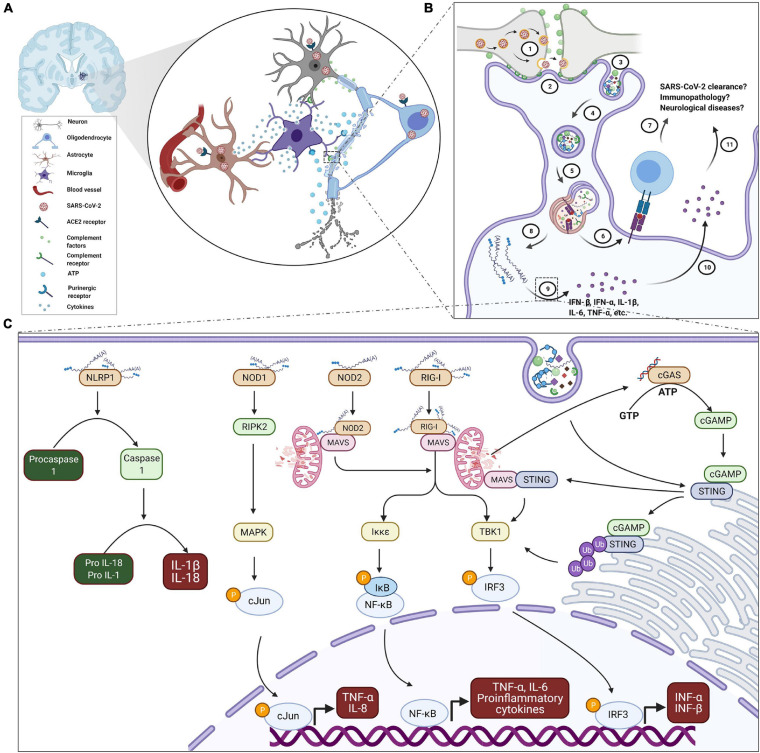
Since December 2019, humankind has been experiencing a ravaging severe acute respiratory syndrome coronavirus 2 (SARS-CoV-2) outbreak, the second coronavirus pandemic in a decade after the Middle East respiratory syndrome coronavirus (MERS-CoV) disease in 2012. Infection with SARS-CoV-2 results in Coronavirus disease 2019 (COVID-19), which is responsible for over 3.1 million deaths worldwide. With the emergence of a second and a third wave of infection across the globe, and the rising record of multiple reinfections and relapses, SARS-CoV-2 infection shows no sign of abating. In addition, it is now evident that SARS-CoV-2 infection presents with neurological symptoms that include early hyposmia, ischemic stroke, meningitis, delirium and falls, even after viral clearance. This may suggest chronic or permanent changes to the neurons, glial cells, and/or brain vasculature in response to SARS-CoV-2 infection or COVID-19. Within the central nervous system (CNS), microglia act as the central housekeepers against altered homeostatic states, including during viral neurotropic infections. In this review, we highlight microglial responses to viral neuroinfections, especially those with a similar genetic composition and route of entry as SARS-CoV-2. As the primary sensor of viral infection in the CNS, we describe the pathogenic and neuroinvasive mechanisms of RNA viruses and SARS-CoV-2 vis-à-vis the microglial means of viral recognition. Responses of microglia which may culminate in viral clearance or immunopathology are also covered. Lastly, we further discuss the implication of SARS-CoV-2 CNS invasion on microglial plasticity and associated long-term neurodegeneration. As such, this review provides insight into some of the mechanisms by which microglia could contribute to the pathophysiology of post-COVID-19 neurological sequelae and disorders, including Parkinson's disease, which could be pervasive in the coming years given the growing numbers of infected and re-infected individuals globally.

Keywords: COVID-19; Parkinson’s disease; SARS-CoV-2; brain; microglia; neurodegenerative diseases; neuropsychiatric disorders; viral RNA neurotropism.


Conflict of interest statement

The authors declare that the research was conducted in the absence of any commercial or financial relationships that could be construed as a potential conflict of interest.

Figures

FIGURE 1
FIGURE 1
Proposed schematic of microglial reactivity and implications in SARS-CoV-2 infection and COVID-19. (A) COVID-19-associated focal hemorrhagic infarcts in the brain are characterized with microglia nodules, degenerating neurons and infiltrated T cells. Thus, microglia may be coordinating the inflammatory events around the infarct’s milieu in a number of ways via reactivity to signals from oligodendrocytes, neurons and astrocytes after SARS-CoV-2 infection, including ATP and complement (C1q or C3) tags, as well as secretion of cytokines. (B) For instance, complement coating of SARS-CoV-2-infected synapses (1) may trigger microglial recruitment and interaction via their complement receptors (2) culminating in encapsulation (3) and phagocytosis (4) of synaptic elements in membrane cargoes, which subsequently fuse with lysosomes for adequate processing (5). In the process, fragments of viral peptides may be presented via MHC-I and/or MHC-II to cytotoxic and/or helper T cells (6), respectively, of elicit adaptive immune response. However excessive phagocytosis of synaptic elements may overwhelm the phagolysosomal processing (8) resulting in the exposure of microglia to SARS-CoV-2 genome and functional/structural impairment of vital organelles. The exposure sensitizes microglia to produce (9) and secrete (10) both antiviral and inflammatory cytokines in significant quantity. Although microglia are equipped with a competent innate recognition system, their contribution in the context of SARS-CoV-2 infection and COVID-19 is yet unknown (7 and 11). (C) For emphasis, upon cytosolic exposure, microglia may detect SARS-CoV-2 genome through a battery of sensors. NLRP1 sensing of dsRNA and ssRNA activates inflammasome, which processes IL-1β and IL-18 production through caspase 1. NOD1 binding of dsRNA activates the translocation of cJun to the nucleus with subsequent upregulation of pro-inflammatory mediators. RIG-I-bound dsRNA and ssRNA as well as NOD-2- ssRNA complex exacerbate production of TNF-α, IL-6, and IL8 through mitochondrial adaptor protein MAVS mediated NF-κB signaling. Simultaneously, they also regulate the transcription of antiviral type 1 interferons through IRF3. In addition, DAMPs from stressed microglial organelles, such as mitochondrial DNA may trigger cGAS receptor to synthesize cGAMP, an agonist of STING. STING activation potentiates IRF3 signaling. Membrane fusion of endosomatic cargoes may also initiate cGAS-independent STING-interferons signaling through MAVS. Thus, characterization of the specific contribution of microglia in the development of neuronal damage and associated neurological sequelae, or the involvement in debris clearance, SARS-CoV-2 resolution and disease outcome is an active area of research. ATP, adenosine triphosphate; COVID-19, coronavirus disease 2019; DAMPs, damage-associated molecular patterns; cGAMP, cyclic GMP-AMP; cGAS, cyclic GMP-AMP synthase; GTP, guanosine triphosphate; IL, interleukin; IκB, inhibitor of κB; Iκκε, IκB kinase; IFN-α/β, interferon alpha/beta; IRF3, interferon Regulatory Factor 3; MAPK, mitogen activated protein kinase; MAVS, mitochondrial antiviral signaling protein; MHC-I/II, major histocompatibility complex I/II; NOD2/1, nucleotide-binding oligomerization domain 2/1; NF-κB, nuclear factor kappa light chain enhancer of activated B cells; NLRP1, NLR family pyrin domain containing 1; P, phosphate; RIG-I, retinoic acid-inducible gene I; RIPK2, receptor interacting serine/threonine protein kinase 2; dsRNA, double stranded viral RNA; ssRNA, single stranded viral RNA; SARS-CoV-2, severe acute respiratory syndrome coronavirus; STING, stimulator of type I interferon genes; TBK-1, TANK-binding kinase 1; TNF-α, tumor necrosis factor alpha; Ub, ubiquitin.

Similar articles

Cited by

References

    1. Adhikari S. P., Meng S., Wu Y. J., Mao Y. P., Ye R. X., Wang Q. Z., et al. (2020). Epidemiology, causes, clinical manifestation and diagnosis, prevention and control of coronavirus disease (COVID-19) during the early outbreak period: a scoping review. Infect. Dis. Poverty 9:29. 10.1186/s40249-020-00646-x - DOI - PMC - PubMed
    1. Aghagoli G., Gallo Marin B., Katchur N. J., Chaves-Sell F., Asaad W. F., Murphy S. A. (2020). Neurological involvement in COVID-19 and potential mechanisms: a review. Neurocrit. Care [Epub ahead of print]. 10.1007/s12028-020-01049-4 - DOI - PMC - PubMed
    1. Agrawal P., Nawadkar R., Ojha H., Kumar J., Sahu A. (2017). Complement evasion strategies of viruses: an overview. Front. Microbiol. 8:1117. 10.3389/fmicb.2017.01117 - DOI - PMC - PubMed
    1. Aguirre S., Maestre A. M., Pagni S., Patel J. R., Savage T., Gutman D., et al. (2012). DENV inhibits type I IFN production in infected cells by cleaving human STING. PLoS Pathog. 8:e1002934. 10.1371/journal.ppat.1002934 - DOI - PMC - PubMed
    1. Aguzzi A., Barres B. A., Bennett M. L. (2013). Microglia: scapegoat, saboteur, or something else? Science 339 156–161. 10.1126/science.1227901 - DOI - PMC - PubMed