. 2021 Jun 15:12:686060.
doi: 10.3389/fimmu.2021.686060. eCollection 2021.

Transcriptomic Analysis and C-Terminal Epitope Tagging Reveal Differential Processing and Signaling of Endogenous TLR3 and TLR7

Affiliations

Transcriptomic Analysis and C-Terminal Epitope Tagging Reveal Differential Processing and Signaling of Endogenous TLR3 and TLR7

Chiung-Ya Chen et al. Front Immunol. .

Abstract

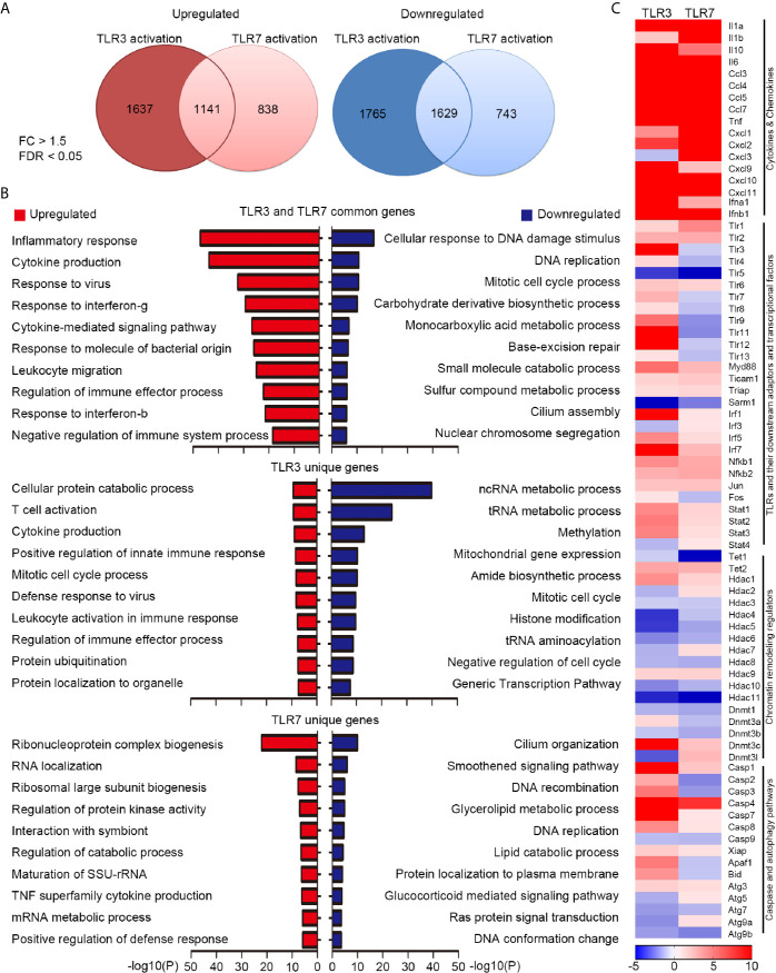
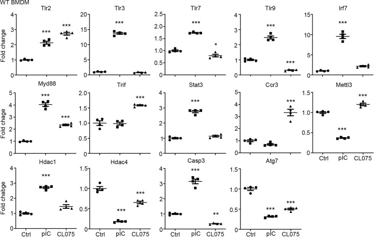
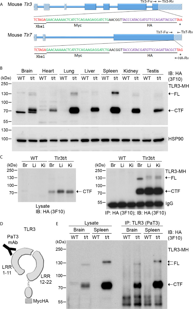
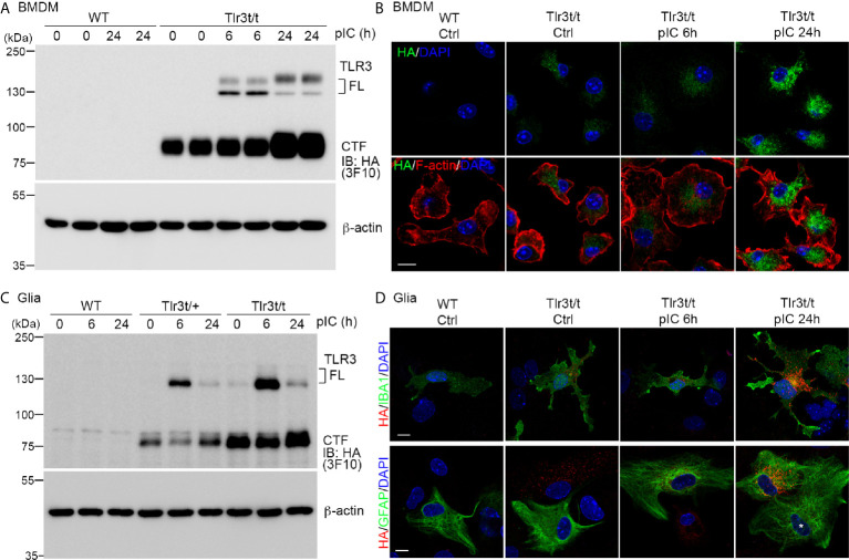
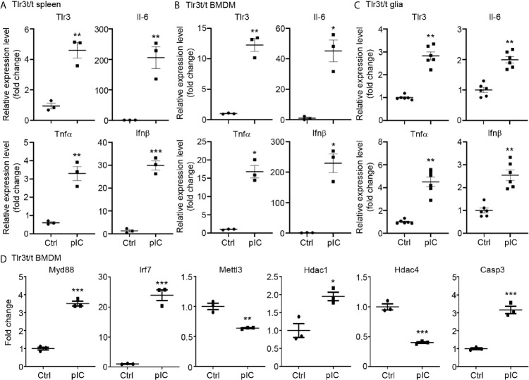
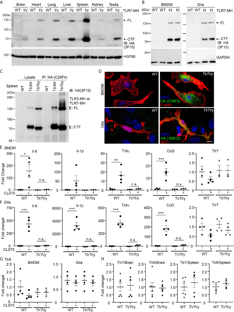
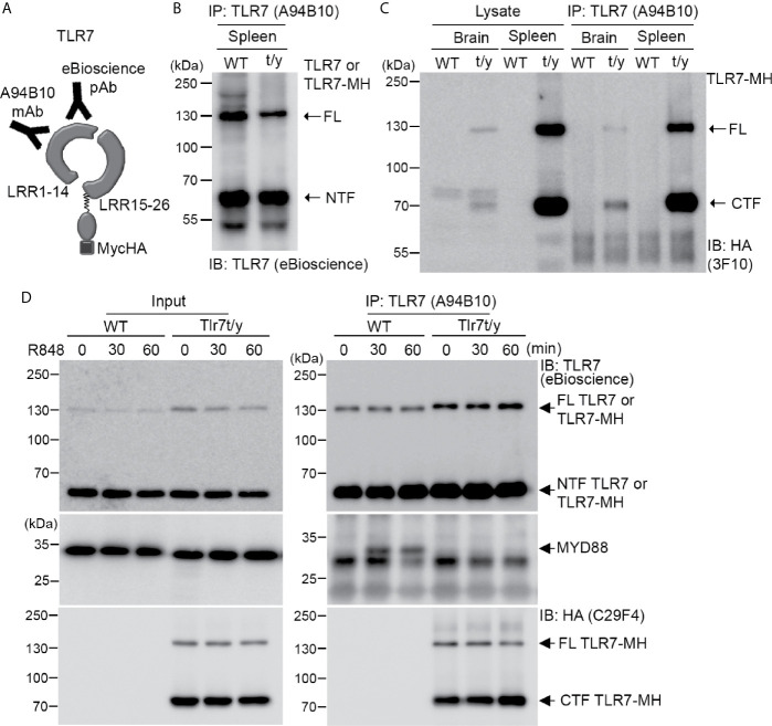
Toll-like receptor (TLR) signaling is critical for defense against pathogenic infection, as well as for modulating tissue development. Activation of different TLRs triggers common inflammatory responses such as cytokine induction. Here, we reveal differential impacts of TLR3 and TLR7 signaling on transcriptomic profiles in bone marrow-derived macrophages (BMDMs). Apart from self-regulation, TLR3, but not TLR7, induced expression of other TLRs, suggesting that TLR3 activation globally enhances innate immunity. Moreover, we observed diverse influences of TLR3 and TLR7 signaling on genes involved in methylation, caspase and autophagy pathways. We compared endogenous TLR3 and TLR7 by using CRISPR/Cas9 technology to knock in a dual Myc-HA tag at the 3' ends of mouse Tlr3 and Tlr7. Using anti-HA antibodies to detect endogenous tagged TLR3 and TLR7, we found that both TLRs display differential tissue expression and posttranslational modifications. C-terminal tagging did not impair TLR3 activity. However, it disrupted the interaction between TLR7 and myeloid differentiation primary response 88 (MYD88), the Tir domain-containing adaptor of TLR7, which blocked its downstream signaling necessary to trigger cytokine and chemokine expression. Our study demonstrates different properties for TLR3 and TLR7, and also provides useful mouse models for further investigation of these two RNA-sensing TLRs.

Keywords: MYD88; RNA-seq; epitope tagging; signalosome; toll-like receptor; transcriptomic analysis.


Conflict of interest statement

The authors declare that the research was conducted in the absence of any commercial or financial relationships that could be construed as a potential conflict of interest.

Figures

Figure 1
Figure 1
Transcriptomic profiling reveals differential downstream targets of TLR3 and TLR7 in BMDMs. (A) Venn diagram showing the overlap and difference between TLR3- and TLR7-regulated genes. Left: upregulated; Right: downregulated. Fold change (FC) > 1.5; false discovery rate (FDR) < 0.05. The numbers of identified genes are also indicated. (B) Gene ontology (GO) of commonly-targeted or uniquely-targeted TLR3 and TLR7 genes based on biological functions. Red bars: upregulated genes; blue bars: downregulated genes. The top ten GOs for each group are shown. (C) Heat map depicting the relative mRNA levels of selected TLR3- and TLR7-regulated genes. Scale bar: z-score.
Figure 2
Figure 2
Quantitative RT-PCR confirms altered gene expression in response to TLR3 or TLR7 activation. Poly(I:C) and CL075 were used to activate TLR3 or TLR7, respectively, in WT BMDMs. The expression levels of indicated genes were determined by quantitative RT-PCR and normalized against internal control Hprt. Data are represented as mean ± SEM (error bars). Each dot indicates the data of one independent experiment. *P < 0.05; **P < 0.01; ***P < 0.001.
Figure 3
Figure 3
Dual tagging of Myc and HA cassettes at the C-terminal ends of the Tlr genes reveals expression and processing of TLR. (A) Schematic of the mouse Tlr3 and Tlr7 genes in which a Myc-HA epitope tag has been inserted before the stop codon. Two primer sets, i.e. Tlr3-Fw and Tlr3-Rv for Tlr3 and Tlr7-Fw, Tlr7-Rv and HA-Rv for Tlr7, were used for genotyping the tagged Tlr3 and Tlr7 mice. The results of genotyping are available in Supplementary Figure 2 . *, stop codon. (B) Detection of TLR3-MH protein in multiple tissues of Tlr3t/t mice using immunoblotting (IB) with anti-HA antibody (3F10). HSP90 was used as a loading control. (C) Anti-HA antibody (3F10) was used in immunoprecipitation (IP) of TLR3-MH from brain (Br), liver (Li) and kidney (Ki) of WT and Tlr3t/t mice. The IP complex was then analyzed by IB with the same anti-HA antibody. (D) Schematic of TLR3 protein with the C-terminal dual Myc-HA tag. The PaT3 monoclonal antibody recognizes the N-terminal region of TLR3. (E) The N-terminal (NTF) and C-terminal (CTF) fragments of TLR3 remain associated with each other after proteolytic cleavage. TLR3 was precipitated using the PaT3 antibody from brain and spleen lysates of WT and Tlr3t/t mice. The IP complex was then subjected to IB analysis using anti-HA antibody (3F10). FL, full-length TLR3-MH; CTF, C-terminal fragment of TLR3-MH; IgG, immunoglobulin heavy chain.
Figure 4
Figure 4
Poly(I:C) stimulation increased endogenous TLR3-MH protein level in Tlr3t/t cells. (A, C) Immunoblot analysis of TLR3 proteins in BMDMs (A) and glia cells (C), as indicated. WT, Tlr3t/+ and Tlr3t/t cells were treated with poly(I:C) or vehicle control. After different incubation times, cell lysates were harvested and analyzed using anti-HA (3F10) and β-actin antibodies (loading control). FL, full-length TLR3; CTF, C-terminal fragment of TLR3. (B, D) Immunofluorescence staining to monitor TLR3 expression in BMDMs (B) and glia cells (D) of WT and Tlr3t/t mice with or without poly(I:C) treatment. Anti-HA antibodies (C29F4 in (B, D) lower panel, or 16B12 in (D) upper panel) were used for dual immunostaining with cell markers. F-actin was used to outline BMDM morphology. IBA1 is a microglia marker. GFAP is a marker for astrocytes. Nuclei were counter-stained with DAPI. Scale bar, 10 μm.
Figure 5
Figure 5
Poly(I:C) stimulation altered the expression of Tlr3 and downstream genes in Tlr3t/t spleen and cells. (A) Quantitative RT-PCR analysis of Tlr3 and the cytokines Il-6, Tnfα and Ifnβ in poly(I:C)-challenged Tlr3t/t mouse spleen. Six hours after an intraperitoneal injection of 5 mg/kg poly(I:C), total RNA was extracted from spleens of Tlr3t/t mice using TRIzol reagent. The expression levels of indicated cytokine genes were normalized against the internal control Hprt. (B–D) Primary cultures of BMDMs (B, D) and glia cells (C) from Tlr3t/t mice were stimulated with 10 μg/ml poly(I:C) for 6 h. The relative RNA levels of indicated genes normalized against the internal control Hprt were analyzed by means of quantitative RT-PCR. Data are represented as mean ± SEM (error bars). Each dot indicates the result of (A) an individual animal or (B–D) an independent culture. *p > 0.05; **p > 0.01; ***p > 0.001.
Figure 6
Figure 6
C-terminal Myc-HA dual tagging results in inactivation of TLR7. (A, B) Immunoblotting using anti-HA antibodies was performed to detect the expression of TLR7-MH in Tlr7t/y tissues (A) and Tlr7t/t cells (B). Different organs and BMDMs and glia cell cultures were examined, as indicated. Samples prepared from WT littermates were used as negative controls. HSP90 and GAPDH were used as a loading control. FL, full-length TLR7-MH; CTF, C-terminal fragment of TLR7-MH. (C) Distinct levels of proteolyzed TLR3-MH and TLR7-MH in mouse spleen. Anti-HA antibody (C29F4) was used to precipitate TLR3-MH and TLR7-MH proteins from Tlr3t/t and Tlr7t/y mouse spleens, respectively. WT mice were used as a negative control. The IP complex was then subjected to IB analysis using anti-HA antibody (3F10). FL: full-length TLR3-MH or TLR7-MH, CTF: C-terminal fragment of TLR3-MH or TLR7-MH. (D) Immunostaining of BMDMs and glial cells using anti-HA and anti-IBA1 (for microglia) antibodies was performed, as indicated. Phalloidin and DAPI were used to label F-actin and nuclei, respectively. Scale bars, 10 μm. (E–G) BMDMs and glia cells prepared from WT and Tlr7t/t mice were treated with 4 μM CL075, a TLR7 agonist, for 6 h. Expression levels of Il-6, Il-1b, Tnfa, Ccl3, Tlr7 and Tlr8 were then determined using quantitative RT-PCR and normalized against the internal control Hprt. (H) Levels of Tlr7 and Tlr8 mRNA are similar between WT and Tlr7t/y mouse organs. Brain and spleen of WT and Tlr7t/y mice were harvested for RNA extraction and then subjected to quantitative RT-PCR to analyze levels of Tlr7 and Tlr8, normalized against the internal control Hprt. Data are represented as mean ± SEM (error bars). Each dot indicates the result of one independent culture. *p > 0.05; **p > 0.01; ***p > 0.001; n.s., non-significant. (E–G) Two-way with Bonferroni’s multiple comparisons test.
Figure 7
Figure 7
Dual C-terminal Myc-HA tagging disrupts the interaction between TLR7 and MYD88. (A) Schematic of TLR7 protein with the dual C-terminal Myc-HA tag. Monoclonal anti-TLR7 antibody (A94B10) and polyclonal TLR7 antibody both target the N-terminal region. (B, C) The N-terminal (NTF) and C-terminal (CTF) fragments of TLR7 remain associated with each other after proteolytic cleavage. TLR7 was immunoprecipitated (IP) using anti-TLR7 antibody (A94B10) from brain and spleen lysates of WT and Tlr7t/y mice. The IP complex was then subjected to immunoblotting (IB) analysis using anti-TLR7 antibody (eBioscience) (B) or anti-HA antibody (3F10) (C). (D) WT and Tlr7t/y BMDMs were treated with a TLR7 agonist (R848) for 30 and 60 min. Cell lysates were then collected and subjected to IP using anti-TLR7 antibody (A94B10). The IP complex was then analyzed by IB using anti-TLR7 (eBioscience) (upper panel), anti-MyD88 (middle panel), and anti-HA (C29F4, lower panel) antibodies. FL, full-length.

References

    1. Akira S, Uematsu S, Takeuchi O. Pathogen Recognition and Innate Immunity. Cell (2006) 124:783–801. 10.1016/j.cell.2006.02.015 - DOI - PubMed
    1. Brubaker SW, Bonham KS, Zanoni I, Kagan JC. Innate Immune Pattern Recognition: A Cell Biological Perspective. Annu Rev Immunol (2015) 33:257–90. 10.1146/annurev-immunol-032414-112240 - DOI - PMC - PubMed
    1. Gay NJ, Gangloff M. Structure and Function of Toll Receptors and Their Ligands. Annu Rev Biochem (2007) 76:141–65. 10.1146/annurev.biochem.76.060305.151318 - DOI - PubMed
    1. Chen CY, Shih YC, Hung YF, Hsueh YP. Beyond Defense: Regulation of Neuronal Morphogenesis and Brain Functions Via Toll-like Receptors. J BioMed Sci (2019) 26:90. 10.1186/s12929-019-0584-z - DOI - PMC - PubMed
    1. Kawai T, Akira S. The Role of Pattern-Recognition Receptors in Innate Immunity: Update on Toll-Like Receptors. Nat Immunol (2010) 11:373–84. 10.1038/ni.1863 - DOI - PubMed

Publication types

MeSH terms