. 2021 May 28;8(3):rbab017.
doi: 10.1093/rb/rbab017. eCollection 2021 Jun.

The biological responses and mechanisms of endothelial cells to magnesium alloy

Affiliations

The biological responses and mechanisms of endothelial cells to magnesium alloy

Zhe Hou et al. Regen Biomater. .

Abstract

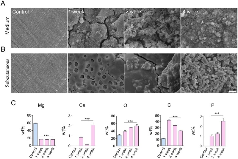
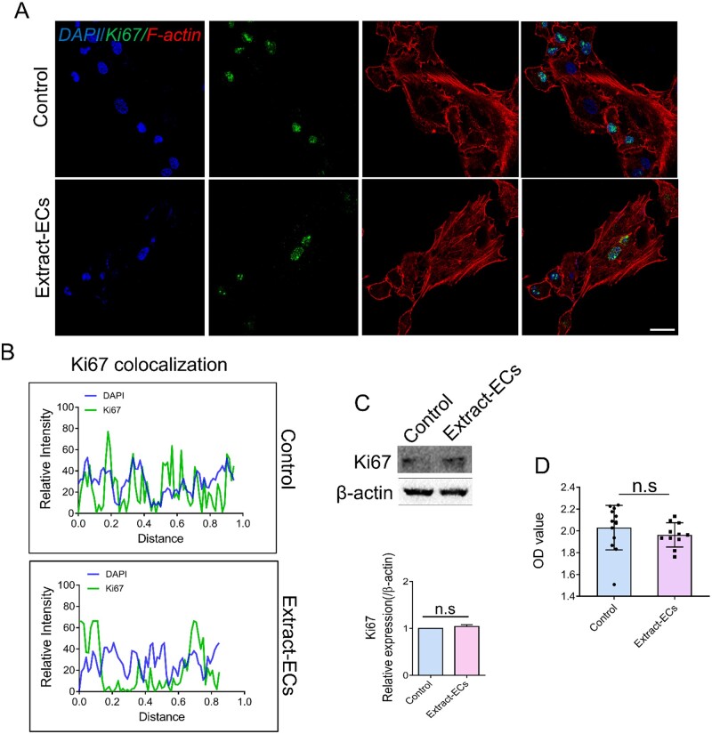
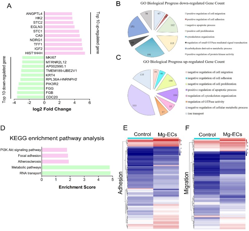
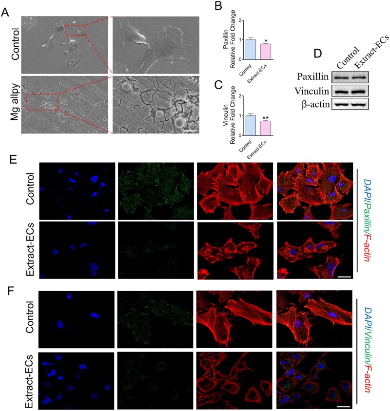
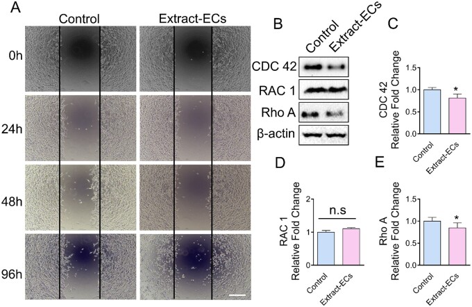
Due to its good biocompatibility and degradability, magnesium alloy (Mg alloy) has shown great promise in cardiovascular stent applications. Rapid stent re-endothelialization is derived from migrated and adhered endothelial cells (ECs), which is an effective way to reduce late thrombosis and inhibit hyperplasia. However, fundamental questions regarding Mg alloy affecting migration and adhesion of ECs are not fully understood. Here, we evaluated the effects of Mg alloy on the ECs proliferation, adhesion and migration. A global gene expression profiling of ECs co-culturing with Mg alloy was conducted, and the adhesion- and migration-related genes were examined. We found that Mg alloy had no adverse effects on ECs viability but significantly affected ECs migration and adhesion. Co-cultured with Mg alloy extract, ECs showed contractive adhesion morphology and decreased motility, which was supported by the down-regulation of adhesion-related genes (Paxillin and Vinculin) and migration-related genes (RAC 1, Rho A and CDC 42). Accordingly, the re-endothelialization of Mg alloy stent was inhibited in vivo. Our results may provide new inspiration for improving the broad application of Mg alloy stents.

Keywords: adhesion; endothelial cells; magnesium alloy; migration.


Figures

Figure 1.
Figure 1.
The degradation of Mg alloy in vivo and in vitro. (A, B) The surface morphologies of degraded discs in medium and subcutaneous tissue at different duration, scale bar = 50 μm. (C) EDS was used to measure the composition of Mg alloy surface elements after implanting in subcutaneous tissue at the indicated time. Data were presented as mean ± SD; statistics were performed using one-way analysis of variance followed by Tukey test, ***P <0.001.
Figure 2.
Figure 2.
The effect of Mg alloy extract on the proliferation of endothelial cells. (A) The distribution and expression of Ki67 in HCAECs with or without Mg alloy extract medium treatment were observed by CLSM. (blue: DAPI; red: F-actin; green: Ki67; scale bar = 10 μm). (B) Co-localization of Ki67 and nuclear was measured by ImageJ software. (C) The protein expression of Ki67 was detected by western blot analysis. β-Actin was used as the internal control (n = 3). (D) The viability of HCACEs was examined by using CCK-8. Data are presented as mean ± SD; statistics were performed by two-tailed unpaired t-test, n.s. denotes not significant.
Figure 3.
Figure 3.
Mg alloy regulated gene expression associated with adhesion and migration. (A) Top 20 significantly changed genes detected by RNA-seq in HCAECs seeded on Mg alloy discs compared with HCAECs (up-regulated coloured red, down-regulated coloured green). (B, C) BP analysis of differently expressed genes in HCAECs seeded on Mg alloy discs compared with HCAECs. (D) KEGG enrichment pathway analysis for changed pathway associated with adhesion and migration in HCAECs seeded on Mg alloy discs compared with HCAECs. (E, F) Differently expressed genes involved in adhesion and migration detected by RNA-seq were shown in the hierarchical diagram in HCAECs seeded on Mg alloy discs compared with HCAECs.
Figure 4.
Figure 4.
The effect of Mg alloy on HCAECs adhesion. (A) SEM images showed the adhesion morphology of HCAECs on the Mg alloy disc. (B, C) The results of qRT-PCR for Paxillin and Vinculin (n = 3). *P <0.05; **P <0.01. β-actin was used as the internal control (n = 3). data are presented as mean ± SD; statistics were performed by two-tailed unpaired t-test. (D) Western blot analysis was used to measure the expression of focal adhesion protein (Paxillin and Vinculin); β-actin was used as the internal control. (E, F) The distribution and expression of Paxillin and Vinculin in HCAECs with or without Mg alloy extract-treatment were detected by immunostaining (blue: DAPI; red: F-actin; green: Paxillin/Vinculin; scale bar = 10 μm).
Figure 5.
Figure 5.
The Effect of Mg alloy extract on HCAECs migration. (A) Wound healing assay shows the difference of cell motility between HCAECs and Extract-ECs at the indicated time, scale bar = 100 μm. (B) The expressions of migration-related proteins (CDC 42, RAC 1 and Rho A) were detected by Western blot. (C–E) The mRNA expressions of CDC 42, RAC 1 and Rho A were examined by qRT-PCR. Data are presented as mean ± SD; statistics were performed by two-tailed unpaired t-test, β-actin was used as the internal control in western blot and qRT-PCR analysis, respectively (n = 3), *P <0.05.
Figure 6.
Figure 6.
The Re-endothelialization condition of the Mg alloy stent after implantation. (A) SEM images showed the re-endothelialization of the stent at different time durations, scale bar = 500 μm (left panel), 200 μm (right panel). (B) EDS images showed the surface chemical element composition change of stents at 2 weeks.

References

    1. Organization WH. Cardiovascular diseases (CVDs): fact sheet 2017. 2017.
    1. Yahagi K, Kolodgie FD, Otsuka F. et al. Pathophysiology of native coronary, vein graft, and in-stent atherosclerosis. Nat Rev Cardiol 2016;13:79–98. - PubMed
    1. Fitzgibbon GM, Kafka HP, Leach AJ. et al. Coronary bypass graft fate and patient outcome: angiographic follow-up of 5,065 grafts related to survival and reoperation in 1,388 patients during 25 years. J Am Coll Cardiol 1996;28:616–26. - PubMed
    1. Neumann F-J, Sousa-Uva M, Ahlsson A. et al. ; ESC Scientific Document Group. 2018 ESC/EACTS Guidelines on myocardial revascularization. Eur Heart J 2019;40:87–165.
    1. Giacoppo D, Alfonso F, Xu B. et al. Drug-coated balloon angioplasty versus drug-eluting stent implantation in patients with coronary stent restenosis. J Am Coll Cardiol 2020;75:2664–78. - PubMed