High-Density of FcγRIIIA+ (CD16+) Tumor-Associated Neutrophils in Metastases Improves the Therapeutic Response of Cetuximab in Metastatic Colorectal Cancer Patients, Independently of the HLA-E/CD94-NKG2A Axis
- PMID: 34211852
- PMCID: PMC8239306
- DOI: 10.3389/fonc.2021.684478
High-Density of FcγRIIIA+ (CD16+) Tumor-Associated Neutrophils in Metastases Improves the Therapeutic Response of Cetuximab in Metastatic Colorectal Cancer Patients, Independently of the HLA-E/CD94-NKG2A Axis
Abstract
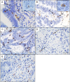
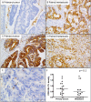
Antibody-dependent cellular cytotoxicity (ADCC) in the anti-tumor effect of cetuximab in metastatic colorectal cancer (mCRC) is only based on the impact of FcγRIIIA (CD16) polymorphisms as predictive of therapeutic response. However, nature, density and therapeutic impact of FcγRIIIA+ (CD16) effector cells in tumor remain poorly documented. Moreover, the inhibition of cetuximab-mediated ADCC induced by NK cells by the engagement of the new inhibitory CD94-NKG2A immune checkpoint has only been demonstrated in vitro. This multicentric study aimed to determine, on paired primary and metastatic tissue samples from a cohort of mCRC patients treated with cetuximab: 1) the nature and density of FcγRIIIA+ (CD16) immune cells, 2) the expression profile of HLA-E/β2m by tumor cells as well as the density of CD94+ immune cells and 3) their impact on both objective response to cetuximab and survival. We demonstrated that FcγRIIIA+ (CD16) intraepithelial immune cells mainly correspond to tumor-associated neutrophils (TAN), and their high density in metastases was significantly associated with a better response to cetuximab, independently of the expression of the CD94/NKG2A inhibitory immune checkpoint. However, HLA-E/β2m, preferentially overexpressed in metastases compared with primary tumors and associated with CD94+ tumor infiltrating lymphocytes (TILs), was associated with a poor overall survival. Altogether, these results strongly support the use of bispecific antibodies directed against both EGFR and FcγRIIIA (CD16) in mCRC patients, to boost cetuximab-mediated ADCC in RAS wild-type mCRC patients. The preferential overexpression of HLA-E/β2m in metastases, associated with CD94+ TILs and responsible for a poor prognosis, provides convincing arguments to inhibit this new immune checkpoint with monalizumab, a humanized anti-NKG2A antibody, in combination with anti- FcγRIIIA/EGFR bispecific antibodies as a promising therapeutic perspective in RAS wild-type mCRC patients.
Keywords: CD16; HLA-E/NKG2A axis; antibody-dependent cellular cytotoxicity; cetuximab; metastatic colorectal carcinoma; tumor-associated neutrophils.
Copyright © 2021 Denis Musquer, Jouand, Pere, Lamer, Bézieau, Matysiak, Faroux, Caroli Bosc, Rousselet, Leclair, Mosnier, Toquet, Gervois and Bossard.
Conflict of interest statement
The authors declare that the research was conducted in the absence of any commercial or financial relationships that could be construed as a potential conflict of interest.
Figures


References
-
- Lanier LL, Le AM, Phillips JH, Babcock GF. Subpopulations of Human Natural Killer Cells Defined by Expression of the Leu-7 (HNK-1) and Leu-11 (NK-15) Antigens. J Immunol (1983) 131:1789–96. - PubMed
LinkOut - more resources
Full Text Sources
Research Materials
Miscellaneous

