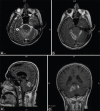
Ruptured Tentorium Originating Masson Tumor
- PMID: 34211895
- PMCID: PMC8202391
- DOI: 10.4103/ajns.AJNS_249_20
Ruptured Tentorium Originating Masson Tumor
Abstract
Intravascular papillary endothelial hyperplasia (IPEH) also known as Masson's tumor, is a benign, slow growing, vascular lesion which is seen very rarely and only a few cases have been reported intracranially in the literature. It has been reported at many sites, but the posterior fossa involvement is very rare. The preoperative diagnosis is very difficult, as there is no enough cases to achieve a clear understanding about the details of its radiological findings. Differential diagnosis have to be made especially from angiosarcoma and meningioma. It is curable by total surgical removal. In this article we presented the characteristic clinical, radiological, perioperative and pathological findings in a case of IPEH in an unusual location, origin and behavior. To best of our knowledge, we presented the first case of IPEH originating from tentorium.
Keywords: Dural attachment; extraaxial lesion; intravascular papillary endothelial hyperplasia; posterior fossa tumor.
Copyright: © 2021 Asian Journal of Neurosurgery.
Conflict of interest statement
There are no conflicts of interest.
Figures


References
-
- Masson M. L'Hemangioendotheliome intravasculaire. Ann Anat Pathol. 1923;93:517.
-
- Shih C, Burgett R, Bonnin J, Boaz J, Ho CY. Intracranial Masson tumor: Case report and literature review. J Neurooncol. 2012;108:211–7. - PubMed
-
- Wang ZH, Hsin CH, Chen SY, Lo CY, Cheng PW. Sinonasal intravascular papillary endothelial hyperplasia successfully treated by endoscopic excision: A case report and review of the literature. Auris Nasus Larynx. 2009;36:363–6. - PubMed
-
- Lee W, Hui Y, Sitoh Y. Intravascular papillary endothelial hyperplasia in an intracranial thrombosed aneurysm 3T magnetic resonance imaging and angiographic features. Singapore Med J. 2004;45:330–3. - PubMed
-
- Miller TR, Mohan S, Tondon R, Montone KT, Palmer JN, Zager EL, et al. Intravascular papillary endothelial hyperplasia of the skull base and intracranial compartment. Clin Neurol Neurosurg. 2013;115:2264–7. - PubMed

