Metabolic Remodeling in Glioma Immune Microenvironment: Intercellular Interactions Distinct From Peripheral Tumors
- PMID: 34211978
- PMCID: PMC8239469
- DOI: 10.3389/fcell.2021.693215
Metabolic Remodeling in Glioma Immune Microenvironment: Intercellular Interactions Distinct From Peripheral Tumors
Abstract
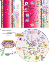
During metabolic reprogramming, glioma cells and their initiating cells efficiently utilized carbohydrates, lipids and amino acids in the hypoxic lesions, which not only ensured sufficient energy for rapid growth and improved the migration to normal brain tissues, but also altered the role of immune cells in tumor microenvironment. Glioma cells secreted interferential metabolites or depriving nutrients to injure the tumor recognition, phagocytosis and lysis of glioma-associated microglia/macrophages (GAMs), cytotoxic T lymphocytes, natural killer cells and dendritic cells, promoted the expansion and infiltration of immunosuppressive regulatory T cells and myeloid-derived suppressor cells, and conferred immune silencing phenotypes on GAMs and dendritic cells. The overexpressed metabolic enzymes also increased the secretion of chemokines to attract neutrophils, regulatory T cells, GAMs, and dendritic cells, while weakening the recruitment of cytotoxic T lymphocytes and natural killer cells, which activated anti-inflammatory and tolerant mechanisms and hindered anti-tumor responses. Therefore, brain-targeted metabolic therapy may improve glioma immunity. This review will clarify the metabolic properties of glioma cells and their interactions with tumor microenvironment immunity, and discuss the application strategies of metabolic therapy in glioma immune silence and escape.
Keywords: glioma; immune escape; metabolic reprogramming; metabolic therapy; tumor microenvironment.
Copyright © 2021 Qiu, Zhong, Li, Li and Fan.
Conflict of interest statement
The authors declare that the research was conducted in the absence of any commercial or financial relationships that could be construed as a potential conflict of interest.
Figures


Similar articles
-
Immune microenvironment of gliomas.Lab Invest. 2017 May;97(5):498-518. doi: 10.1038/labinvest.2017.19. Epub 2017 Mar 13. Lab Invest. 2017. PMID: 28287634 Review.
-
Metabolic and functional reprogramming of myeloid-derived suppressor cells and their therapeutic control in glioblastoma.Cell Stress. 2019 Jan 23;3(2):47-65. doi: 10.15698/cst2019.02.176. Cell Stress. 2019. PMID: 31225500 Free PMC article. Review.
-
Origin, activation, and targeted therapy of glioma-associated macrophages.Front Immunol. 2022 Oct 6;13:974996. doi: 10.3389/fimmu.2022.974996. eCollection 2022. Front Immunol. 2022. PMID: 36275720 Free PMC article. Review.
-
Role of myeloid cells in the immunosuppressive microenvironment in gliomas.Immunobiology. 2020 Jan;225(1):151853. doi: 10.1016/j.imbio.2019.10.002. Epub 2019 Oct 19. Immunobiology. 2020. PMID: 31703822 Review.
-
Mechanisms of intimate and long-distance cross-talk between glioma and myeloid cells: how to break a vicious cycle.Biochim Biophys Acta. 2014 Dec;1846(2):560-75. doi: 10.1016/j.bbcan.2014.10.003. Epub 2014 Oct 20. Biochim Biophys Acta. 2014. PMID: 25453365 Review.
Cited by
-
Ginsenosides Rg1 and CK Control Temozolomide Resistance in Glioblastoma Cells by Modulating Cholesterol Efflux and Lipid Raft Distribution.Evid Based Complement Alternat Med. 2022 Oct 10;2022:1897508. doi: 10.1155/2022/1897508. eCollection 2022. Evid Based Complement Alternat Med. 2022. PMID: 36276866 Free PMC article.
-
Heterogeneity of glioblastoma stem cells in the context of the immune microenvironment and geospatial organization.Front Oncol. 2022 Oct 19;12:1022716. doi: 10.3389/fonc.2022.1022716. eCollection 2022. Front Oncol. 2022. PMID: 36338705 Free PMC article. Review.
-
Metabolism: an important player in glioma survival and development.Discov Oncol. 2024 Oct 22;15(1):577. doi: 10.1007/s12672-024-01402-5. Discov Oncol. 2024. PMID: 39436434 Free PMC article. Review.
-
Lactate dehydrogenase A regulates tumor-macrophage symbiosis to promote glioblastoma progression.Nat Commun. 2024 Mar 5;15(1):1987. doi: 10.1038/s41467-024-46193-z. Nat Commun. 2024. PMID: 38443336 Free PMC article.
-
Changes in Brain Neuroimmunology Following Injury and Disease.Front Integr Neurosci. 2022 Apr 27;16:894500. doi: 10.3389/fnint.2022.894500. eCollection 2022. Front Integr Neurosci. 2022. PMID: 35573444 Free PMC article. Review.
References
-
- Alban T. J., Alvarado A. G., Sorensen M. D., Bayik D., Volovetz J., Serbinowski E., et al. (2018). Global immune fingerprinting in glioblastoma patient peripheral blood reveals immune-suppression signatures associated with prognosis. JCI Insight 3:e122264. 10.1172/jci.insight.122264 - DOI - PMC - PubMed
Publication types
LinkOut - more resources
Full Text Sources

