Review
. 2021 Sep;473(9):1437-1454.
doi: 10.1007/s00424-021-02570-x. Epub 2021 Jul 1.

Cav1.4 dysfunction and congenital stationary night blindness type 2

Affiliations
Review

Cav1.4 dysfunction and congenital stationary night blindness type 2

Alexandra Koschak et al. Pflugers Arch. 2021 Sep.

Abstract

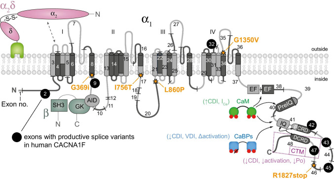
Cav1.4 L-type Ca2+ channels are predominantly expressed in retinal neurons, particularly at the photoreceptor terminals where they mediate sustained Ca2+ entry needed for continuous neurotransmitter release at their ribbon synapses. Cav1.4 channel gating properties are controlled by accessory subunits, associated regulatory proteins, and also alternative splicing. In humans, mutations in the CACNA1F gene encoding for Cav1.4 channels are associated with X-linked retinal disorders such as congenital stationary night blindness type 2. Mutations in the Cav1.4 protein result in a spectrum of altered functional channel activity. Several mouse models broadened our understanding of the role of Cav1.4 channels not only as Ca2+ source at retinal synapses but also as synaptic organizers. In this review, we highlight different structural and functional phenotypes of Cav1.4 mutations that might also occur in patients with congenital stationary night blindness type 2. A further important yet mostly neglected aspect that we discuss is the influence of alternative splicing on channel dysfunction. We conclude that currently available functional phenotyping strategies should be refined and summarize potential specific therapeutic options for patients carrying Cav1.4 mutations. Importantly, the development of new therapeutic approaches will permit a deeper understanding of not only the disease pathophysiology but also the physiological function of Cav1.4 channels in the retina.

Keywords: Calcium channel; Cav1.4; Channel modulation; Channelopathy; Congenital stationary night blindness type 2; Retinal disease.


Conflict of interest statement

The authors declare no competing interests.

Figures

Fig. 1
Fig. 1
Protein topology of human Cav1.4 with coding exons, alternatively spliced exons (see Table 2) and selected mutations annotated (according to human reference sequence NP_005174) together with an intracellular β and an extracellular α2δ and potential interaction sites with calmodulin (CaM) and Ca2+ binding proteins (CaBP). Abbreviations: VDI, voltage-dependence of inactivation, CDI, Ca2+-dependence of inactivation; Po, open probability, AID, alpha-interaction domain; EF, EF-hand motif; other abbreviations, see main text
Fig. 2
Fig. 2
Dual role of Cav1.4 channels in the photoreceptor synapse. Presynaptic Cav1.4 channels serve as sources of Ca2+ ions and play a role as synaptic organizer protein. Ca2+ influx (ICa) changes according to the Cav1.4 mutation (KO, loss-of-function [58]; G369i, non-conducting [61]; I756T, gain-of-function [35]) and exerts different structural effects: left: the presence of sprouting second order neurons (BC, bipolar cell; HC, horizontal cell) and right: changes in the ribbon structure. The inset shows the lateral view on mature (horseshoe-shaped) and immature (round or elongated) ribbons. In knock-out and I756T retinas synaptic terminals were not only located in the outer plexiform layer (OPL) but were also displaced in the outer nuclear layer (ONL) [51, 58, 120]
Fig. 3
Fig. 3
Cav1.4 mutations lead to a change in the Ca2+ dynamics within the photoreceptor voltage range. The current density of different Cav1.4 CSNB2-mutation relative to wild-type Cav1.4 channels (Cav1.4-WT, green) is indicated. Ca2+ currents measured in rod photoreceptors are the reference for the voltage dependence of Cav1.4 wild-type currents ([5]). Current densities were taken from literature: wild type: open circles, 2 mM Ca2+ [20] (set as − 1); I756T: filled circles, 5 mM Ca2+ [35]; L860P: diamonds, 15 mM Ca2+ [20]; R1827stop: stars, 2 mM Ca2+ [20]; CaBP KO: triangles, 20 mM Ca2+[72]. The photoreceptor voltage range is highlighted in orange: from ~  − 60 mV upon light on to ~  − 35 mV at light off. The inset shows currents that were baseline corrected to highlight the change in Ca2+ influx within the photoreceptor voltage range
Fig. 4
Fig. 4
Structure model of the wild type and the G1350V mutant Cav1.4 α1 subunit. Panel a: top view of the Cav1.4 α1 subunit, highlighting the position of the G1350V mutation, which is located at the end of the IVS5 transmembrane helix. Panels b and c illustrate the interdomain interaction of VSD III (K966) with the pore loop of VSD IV (G1350) the wildtype Cav1.4 α1 subunit which cannot be formed between V1350 and K966 (d, e)

References

    1. Abdelkader E, AlHilali S, Neuhaus C, Bergmann C, AlMurshed T, Schatz P. Congenital stationary night blindness associated with morning glory disc malformation: a novel hemizygous mutation in CACNA1F. Ophthalmic Genet. 2018;39:659–661. doi: 10.1080/13816810.2018.1498526. - DOI - PubMed
    1. Aboshiha J, Dubis AM, Carroll J, Hardcastle AJ, Michaelides M. The cone dysfunction syndromes. Br J Ophthalmol. 2016;100:115–121. doi: 10.1136/bjophthalmol-2014-306505. - DOI - PMC - PubMed
    1. Akopian A, Johnson J, Gabriel R, Brecha N, Witkovsky P. Somatostatin modulates voltage-gated K(+) and Ca2+ currents in rod and cone photoreceptors of the salamander retina. J Neurosci. 2000;20:929–936. doi: 10.1523/JNEUROSCI.20-03-00929.2000. - DOI - PMC - PubMed
    1. Audo I, Bujakowska KM, Leveillard T, Mohand-Said S, Lancelot ME, Germain A, Antonio A, Michiels C, Saraiva JP, Letexier M, Sahel JA, Bhattacharya SS, Zeitz C. Development and application of a next-generation-sequencing (NGS) approach to detect known and novel gene defects underlying retinal diseases. Orphanet J Rare Dis. 2012;7:8. doi: 10.1186/1750-1172-7-8. - DOI - PMC - PubMed
    1. Babai N, Thoreson WB. Horizontal cell feedback regulates calcium currents and intracellular calcium levels in rod photoreceptors of salamander and mouse retina. J Physiol. 2009;587:2353–2364. doi: 10.1113/jphysiol.2009.169656. - DOI - PMC - PubMed

Publication types

MeSH terms

Substances

Supplementary concepts