

Endoscopic mucosal resection of a large inflammatory fibroid polyp (Vanek's tumor): a case report
- PMID: 34212909
- PMCID: PMC8343751
- DOI: 10.23750/abm.v92i3.11317
Endoscopic mucosal resection of a large inflammatory fibroid polyp (Vanek's tumor): a case report
Abstract
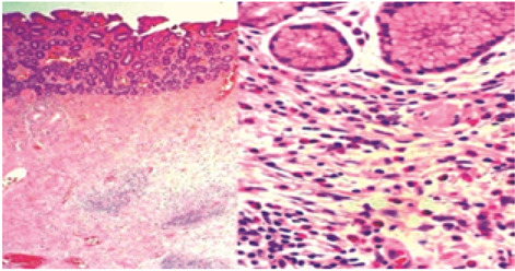
Vanek's Tumor (inflammatory fibroid polyp) is a rare benign mesenchymal lesion occurring throughout the digestive tract. Classical Vanek's tumor ("gastric") contains concentric formations of proliferating spindle cells, which are CD34 positive. Atypical-inflammatory pseudotumor-like Vanek's tumor ("intestinal") lacks concentric formations and is CD34 negative. A 70-years-old man patient presented during hematochemical routine tests, sideropenic anemia and leukopiastrinosis. The patient performed osteomyelitis biopsy and esophagogastroduodenoscopy (EGD) showing a gastric wall with nodular appearance and, in antrum pre-pyloric, a polypoid pedunculated lesion, measuring approximately 3 cm in diameter, surrounded by hyperemic mucosa. The lesion then was removed by en bloc endoscopic mucosal resection (EMR) and histo-morphological, immune-cytochemical and biomolecular evaluations were performed. The data were compatible with a benign polyp fibroid inflammatory (Vanek's Tumor). The results of this study suggest that endoscopic mucosal resection is a safe and efficacy solution for the resection of these gastrointestinal polyps and the two morphological patterns of Vanek's tumor more probably represent only variants of one type of tumor than two different lesions. BRAF mutations were not shown growth PDGFRA wild-type Vanek's tumor.
Conflict of interest statement
Each author declares that he or she has no commercial associations (e.g. consultancies, stock ownership, equity interest, patent/licensing arrangement etc.) that might pose a conflict of interest in connection with the submitted article.
Figures

References
-
- Helwig EB, Ranier A. Inflammatory fibroid polyps of the stomach. Surg Gynecol Obstet. 1953;96:335–67. - PubMed
-
- Barussaud M, Regenet N, Briennon X, et al. Clinical Spectrum and Surgical Approach of Adult Intussusceptions: A Multicentric Study. Int J Colorectal Dis. 2006;21(8):834–9. - PubMed
-
- Nishiyama Y, Koyama S, Andoh A, et al. Gastric inflammatory fibroid polyp treated with Helicobacter pylori eradication therapy. Intern Med. 2003;42:263–7. - PubMed
-
- Shalom A, Wasserman I, Segal M, Orda R. Inflammatory fibroid polyp and Helicobacter pylori. Aetiology or coincidence? Eur J Surg. 2000;166:54–7. - PubMed
Publication types
MeSH terms
LinkOut - more resources
Full Text Sources
Research Materials
Miscellaneous
