

Application of EUS-based techniques in the evaluation of pancreatic cystic neoplasms
- PMID: 34213426
- PMCID: PMC8411565
- DOI: 10.4103/EUS-D-20-00216
Application of EUS-based techniques in the evaluation of pancreatic cystic neoplasms
Abstract
Pancreatic cystic neoplasms (PCNs) are being detected increasingly frequently due to the widespread use of high-resolution abdominal imaging modalities. Some subtypes of PCNs have the potential for malignant transformation. Therefore, accurate diagnosis of PCNs is crucial to determine whether surgical resection or surveillance is the best management strategy. However, the current cross-section imaging modalities are not accurate enough to enable definite diagnoses. In the last decade, EUS-based techniques have emerged, aiming to overcome the limitations of standard cross-section imaging modalities. These novel EUS-based techniques were primarily designed to acquire distinct images to make radiological diagnoses, collect cyst fluid to undergo biochemical or molecular analyses, and obtain tissue to conclude the pathological diagnoses. In this article, we present a comprehensive and critical review of these emerging EUS techniques for the diagnosis of PCNs, with emphasis being placed on the advantages, feasibilities, diagnostic performances, and limitations of these novel techniques.
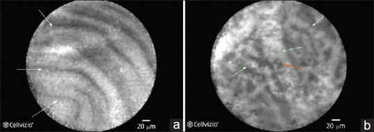
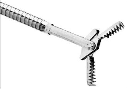
Keywords: EUS; biopsy; confocal laser endomicroscopy; contrast-enhanced harmonic; cyst fluid analysis; cystoscopy; pancreatic cystic neoplasm.
Conflict of interest statement
None
Figures
Similar articles
-
Factors affecting the diagnostic value of liquid-based cytology by EUS-FNA in the diagnosis of pancreatic cystic neoplasms.Endosc Ultrasound. 2024 Mar-Apr;13(2):94-99. doi: 10.1097/eus.0000000000000041. Epub 2023 Dec 20. Endosc Ultrasound. 2024. PMID: 38947751 Free PMC article.
-
New horizons in the endoscopic ultrasonography-based diagnosis of pancreatic cystic lesions.World J Gastroenterol. 2018 Jul 14;24(26):2853-2866. doi: 10.3748/wjg.v24.i26.2853. World J Gastroenterol. 2018. PMID: 30018480 Free PMC article. Review.
-
Diagnosis of pancreatic cysts: EUS-guided, through-the-needle confocal laser-induced endomicroscopy and cystoscopy trial: DETECT study.Gastrointest Endosc. 2015 May;81(5):1204-14. doi: 10.1016/j.gie.2014.10.025. Epub 2015 Jan 26. Gastrointest Endosc. 2015. PMID: 25634486 Clinical Trial.
-
The Role of Endoscopic Ultrasound in the Diagnosis of Cystic Lesions of the Pancreas.Visc Med. 2018 Jul;34(3):192-196. doi: 10.1159/000489242. Epub 2018 Jun 8. Visc Med. 2018. PMID: 30140684 Free PMC article. Review.
-
Age and cystic size are associated with clinical impact of endoscopic ultrasonography-guided fine-needle aspiration on the management of pancreatic cystic neoplasms.Scand J Gastroenterol. 2019 Apr;54(4):506-512. doi: 10.1080/00365521.2019.1601254. Epub 2019 Apr 12. Scand J Gastroenterol. 2019. PMID: 30978145
Cited by
-
Factors affecting the diagnostic value of liquid-based cytology by EUS-FNA in the diagnosis of pancreatic cystic neoplasms.Endosc Ultrasound. 2024 Mar-Apr;13(2):94-99. doi: 10.1097/eus.0000000000000041. Epub 2023 Dec 20. Endosc Ultrasound. 2024. PMID: 38947751 Free PMC article.
-
Threshold of Main Pancreatic Duct Diameter in Identifying Malignant Intraductal Papillary Mucinous Neoplasm by Magnetic Resonance Imaging.Technol Cancer Res Treat. 2023 Jan-Dec;22:15330338231170942. doi: 10.1177/15330338231170942. Technol Cancer Res Treat. 2023. PMID: 37078135 Free PMC article.
-
Basic Principles and Role of Endoscopic Ultrasound in Diagnosis and Differentiation of Pancreatic Cancer from Other Pancreatic Lesions: A Comprehensive Review of Endoscopic Ultrasound for Pancreatic Cancer.J Clin Med. 2024 Apr 28;13(9):2599. doi: 10.3390/jcm13092599. J Clin Med. 2024. PMID: 38731128 Free PMC article. Review.
-
The Latest Advancements in Diagnostic Role of Endosonography of Pancreatic Lesions.J Clin Med. 2023 Jul 12;12(14):4630. doi: 10.3390/jcm12144630. J Clin Med. 2023. PMID: 37510744 Free PMC article. Review.
-
The clinical impact of endoscopic ultrasound-guided fine-needle aspiration on the patients with low-risk pancreatic cystic lesions.Front Oncol. 2022 Aug 5;12:961293. doi: 10.3389/fonc.2022.961293. eCollection 2022. Front Oncol. 2022. PMID: 35992791 Free PMC article.
References
-
- Sun L, Wang Y, Jiang F, et al. Prevalence of pancreatic cystic lesions detected by magnetic resonance imaging in the Chinese population. J Gastroenterol Hepatol. 2019;34:1656–62. - PubMed
-
- Zerboni G, Signoretti M, Crippa S, et al. Systematic review and meta-analysis: Prevalence of incidentally detected pancreatic cystic lesions in asymptomatic individuals. Pancreatology. 2019;19:2–9. - PubMed
-
- Kromrey ML, Bülow R, Hübner J, et al. Prospective study on the incidence, prevalence and 5-year pancreatic-related mortality of pancreatic cysts in a population-based study. Gut. 2018;67:138–45. - PubMed
-
- Brugge WR, Lauwers GY, Sahani D, et al. Cystic neoplasms of the pancreas. N Engl J Med. 2004;351:1218–26. - PubMed
-
- Elta GH, Enestvedt BK, Sauer BG, et al. ACG clinical guideline: Diagnosis and management of pancreatic cysts. Am J Gastroenterol. 2018;113:464–79. - PubMed
