. 2021 Aug;25(15):7342-7353.
doi: 10.1111/jcmm.16763. Epub 2021 Jul 2.

The molecular mechanism underlying mitophagy-mediated hippocampal neuron apoptosis in diabetes-related depression

Affiliations

The molecular mechanism underlying mitophagy-mediated hippocampal neuron apoptosis in diabetes-related depression

Jian Liu et al. J Cell Mol Med. 2021 Aug.

Abstract

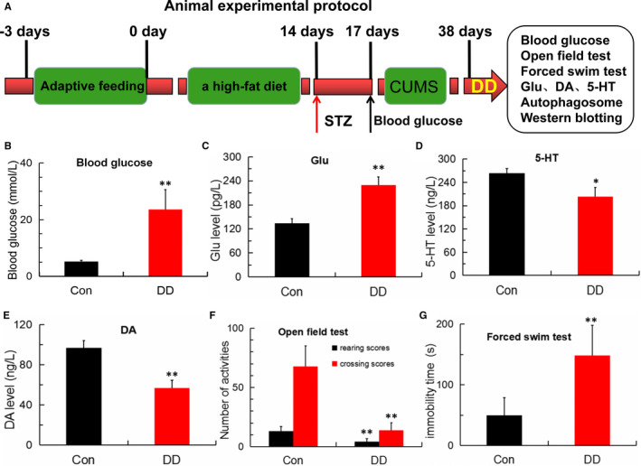
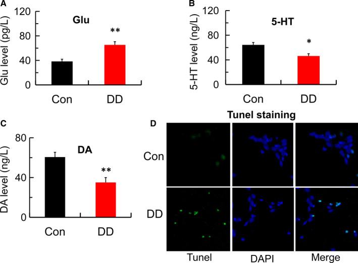
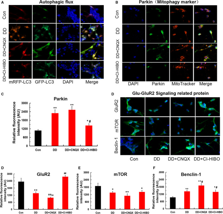
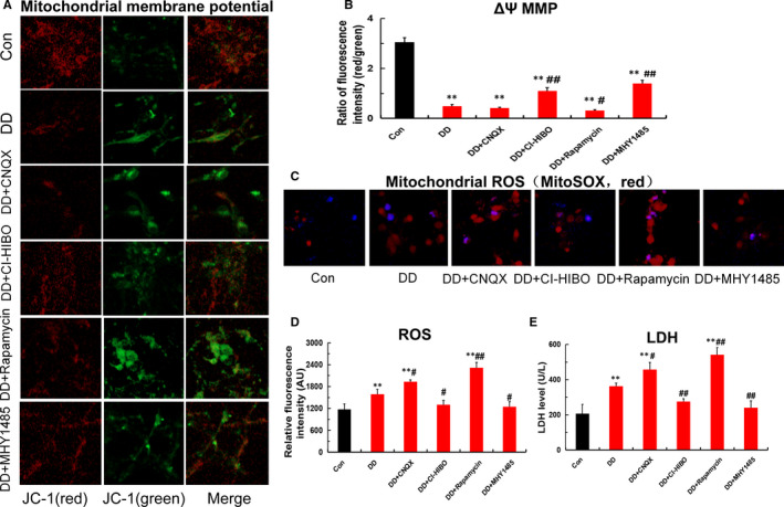
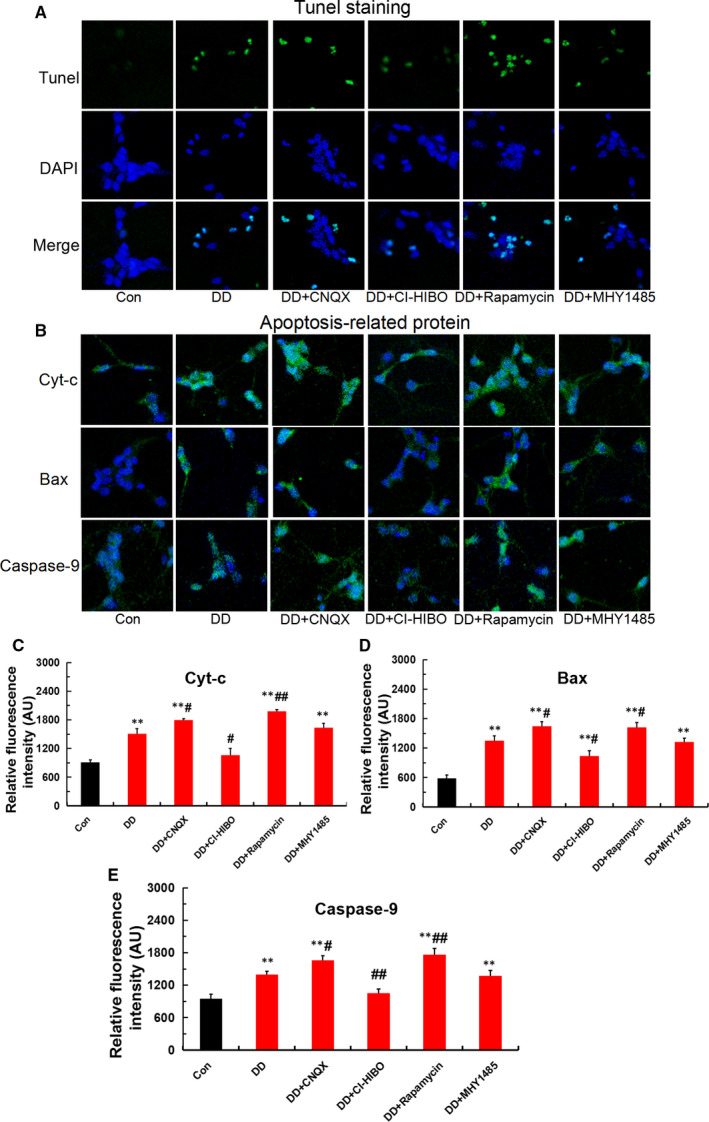
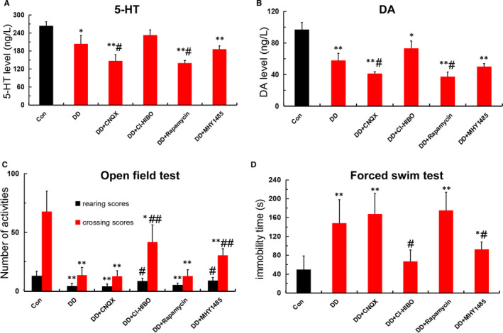
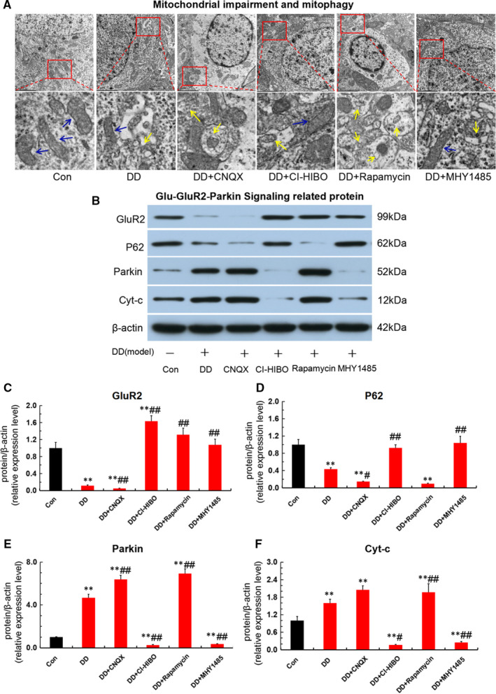
Diabetes-related depression (DD) is a major complication of diabetes mellitus. Our previous studies indicated that glutamate (Glu) and hippocampal neuron apoptosis are key signal and direct factor leading to diabetes-related depression, respectively. However, the accurate pathogenesis remains to be unclear. We hypothesized that diabetes-related depression might be associated with the mitophagy-mediated hippocampal neuron apoptosis, triggered by aberrant Glu-glutamate receptor2 (GluR2)-Parkin pathway. To testify this hypothesis, here the rat model of DD in vivo and in vitro were both established so as to uncover the potential mechanism of DD based on mitophagy and apoptosis. We found that DD rats exhibit an elevated glutamate levels followed by monoamine neurotransmitter deficiency and depressive-like behaviour, and DD modelling promoted autophagosome formation and caused mitochondrial impairment, eventually leading to hippocampal neuron apoptosis via aberrant Glu-GluR2-Parkin pathway. Further, in vitro study demonstrated that the simulated DD conditions resulted in an abnormal glutamate and monoamine neurotransmitter levels followed by autophagic flux increment, mitochondrial membrane potential reduction and mitochondrial reactive oxygen species and lactic dehydrogenase elevation. Interestingly, both GluR2 and mammalian target of rapamycin (mTOR) receptor blocker aggravated mitophagy-induced hippocampal neuron apoptosis and abnormal expression of apoptotic protein. In contrast, both GluR2 and mTOR receptor agonist ameliorated those apoptosis in simulated DD conditions. Our findings revealed that mitophagy-mediated hippocampal neuron apoptosis, triggered by aberrant Glu-GluR2-Parkin pathway, is responsible for depressive-like behaviour and monoamine neurotransmitter deficiency in DD rats. This work provides promising molecular targets and strategy for the treatment of DD.

Keywords: Apoptosis; Diabetes-related depression; GluR2; Glutamate; Hippocampal neuron; Mitophagy; Parkin.


Conflict of interest statement

All authors declare that they have no competing interests.

Figures

FIGURE 1
FIGURE 1
Abnormal Glu, 5‐HT, DA levels and depressive‐like behaviour in DD rats. (A) Experimental protocol. Rats were adaptively fed for 3 days then given to a high‐fat diet for 2 weeks and injected with STZ and followed by subjected CUMS for 21 days to establish the DD model. (B) Blood‐glucose levels detected by a blood‐glucose sensor in control and DD model group (n = 5; **P < .001 vs Con). (C) Glutamate content detected by ELISA in control and DD model group (n = 4; **P < .001 vs Con). (D‐E) 5‐HT and DA levels detected by ELISA in control and DD model group (n = 4; *P < .05, **P < .001 vs Con). (F‐G) Number of activities in the open‐field test and the immobility time in the forced swimming test (n = 5; **P < .001 vs Con)
FIGURE 2
FIGURE 2
Abnormal Glu, 5‐HT, DA levels and hippocampal neurons apoptosis in simulated DD conditions. (A) Glutamate content detected by ELISA in control and DD model group (n = 4; **P < .001 vs Con). (B‐C) 5‐HT and DA levels detected by ELISA in control and DD model group (n = 4; *P < .05, **P < .001 vs Con). (D) Hippocampal neurons apoptosis detected by Tunel staining in control and DD model group (magnification:200×)
FIGURE 3
FIGURE 3
The simulated DD conditions lead to hippocampal neurons mitophagy via Glu‐GluR2 signal. (A) Autophagic flux was monitored by confocal microscopy and mRFP‐GFP‐LC3 virus transfection. (B‐C) Mitophagy marker protein of Parkin detected by immunofluorescence staining in each group (magnification:200×; n = 3; *P < .05, **P < .001 vs Con; # P < .05 vs DD). (D‐G) Glu‐GluR2 signal‐related proteins of GluR2, mTOR and Beclin‐1 detected by immunofluorescence staining in each group (magnification:200×; n = 3; *P < .05, **P < .001vs Con; # P < .05 vs DD, ## P < .01 vs DD)
FIGURE 4
FIGURE 4
Mitochondrial dysfunction in hippocampal neurons under the simulated DD conditions. (A‐B) Mitochondrial membrane potential (MMP) detected by JC‐1 fluorescent probe in each group(magnification:200×; n = 3; **P < .001 vs Con; # P < .05, ## P < .01 vs DD). (C‐D) Mitochondrial ROS detected by MitoSOX probe in each group (magnification:200×; n = 3; **P < .001 vs Con; # P < .05, ## P < .01 vs DD). (E) LDH detected by ELISA in each group (n = 3; **P < .001vs Con; # P < .05, ## P < .01 vs DD)
FIGURE 5
FIGURE 5
Mitophagy‐induced hippocampal neurons apoptosis under the simulated DD conditions. (A) Hippocampal neurons apoptosis detected by Tunel staining in each group(magnification:200×). (B‐E) Apoptotic proteins of Cyt‐c, Bax and caspase‐9 detected by immunofluorescence staining in each group (magnification:200×; n = 3; **P <.001 vs Con; # P < .05, ## P < .01 vs DD)
FIGURE 6
FIGURE 6
Aberrant Glu‐GluR2‐Parkin pathway leads to monoamine neurotransmitter deficiency and depressive‐like behaviour in DD rats. (A‐B) 5‐HT and DA levels detected by ELISA in each group (n = 4; *P < .05,**P < .001vs Con; # P < .05, ## P < .01 vs DD). (C‐D)Number of activities in open‐field test and the immobility time in forced swimming test among each group (n = 5; *P < .05, **P < .001 vs Con; # P < .05 vs DD)
FIGURE 7
FIGURE 7
Aberrant Glu‐GluR2‐Parkin pathway triggered mitophagy‐mediated hippocampal neurons apoptosis in DD rats. (A)Mitophagy and autophagosome detected by transmission electron microscope in each group. The ‘blue arrow’ represents for the normal mitochondria and the ‘yellow arrow’ shows the mitophagy. (B‐F)Expression of Glu‐GluR2‐Parkin pathway protein and endogenous apoptotic protein for Cyt‐c detected by Western blot assay in each group (n = 3; **P < .001vs Con; # P < .05, ## P < .01 vs DD)

References

    1. Rami‐Merhar B, Fröhlich‐Reiterer E, Hofer SE. Diabetes mellitus in childhood and adolescence (Update 2019). Wien Klin Wochenschr. 2019;131(Suppl 1):85‐90. - PubMed
    1. Cole JB, Florez JC. Genetics of diabetes mellitus and diabetes complications. Nat Rev Nephrol. 2020;16(7):377‐390. - PMC - PubMed
    1. Sartorius N. Depression and diabetes. Dialogues Clin Neurosci. 2018;20(1):47‐52. - PMC - PubMed
    1. Lloyd CE, Nouwen A, Sartorius N et al. Prevalence and correlates of depressive disorders in people with type 2 diabetes: results from the international prevalence and treatment of diabetes and depression (INTERPRET‐DD) study, a collaborative study carried out in 14 countries. Diabet Med. 2018;35(6):760‐769. - PubMed
    1. Semenkovich K, Brown ME, Svrakic DM, Lustman PJ. Depression in type 2 diabetes mellitus: prevalence, impact, and treatment. Drugs. 2015;75(6):577‐587. - PubMed

Publication types

MeSH terms