Review
. 2021 Dec:71:27-35.
doi: 10.1016/j.sbi.2021.05.010. Epub 2021 Jun 30.

Structural perspectives on H2S homeostasis

Affiliations
Review

Structural perspectives on H2S homeostasis

Aaron P Landry et al. Curr Opin Struct Biol. 2021 Dec.

Abstract

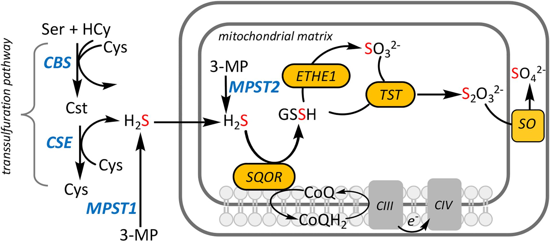
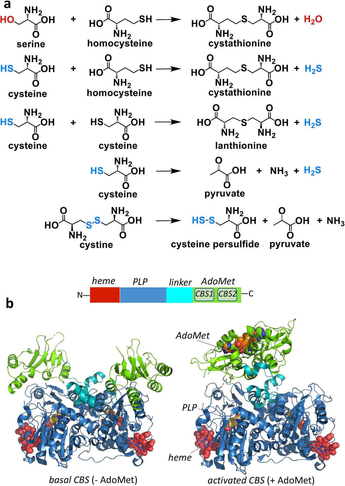
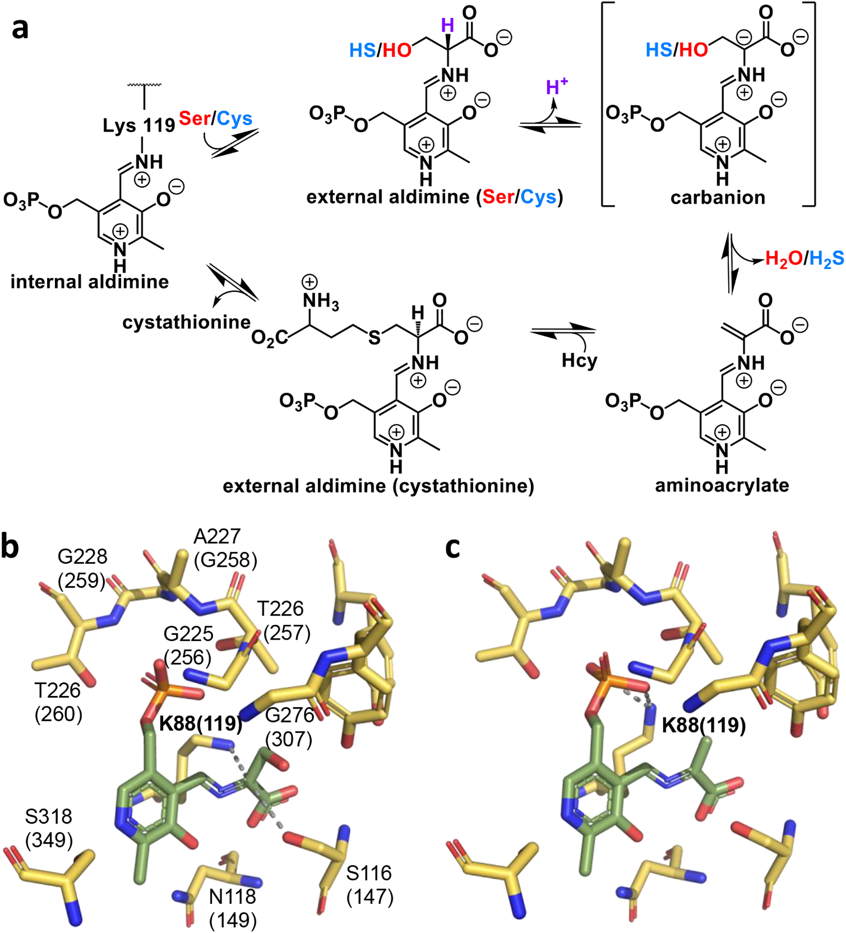
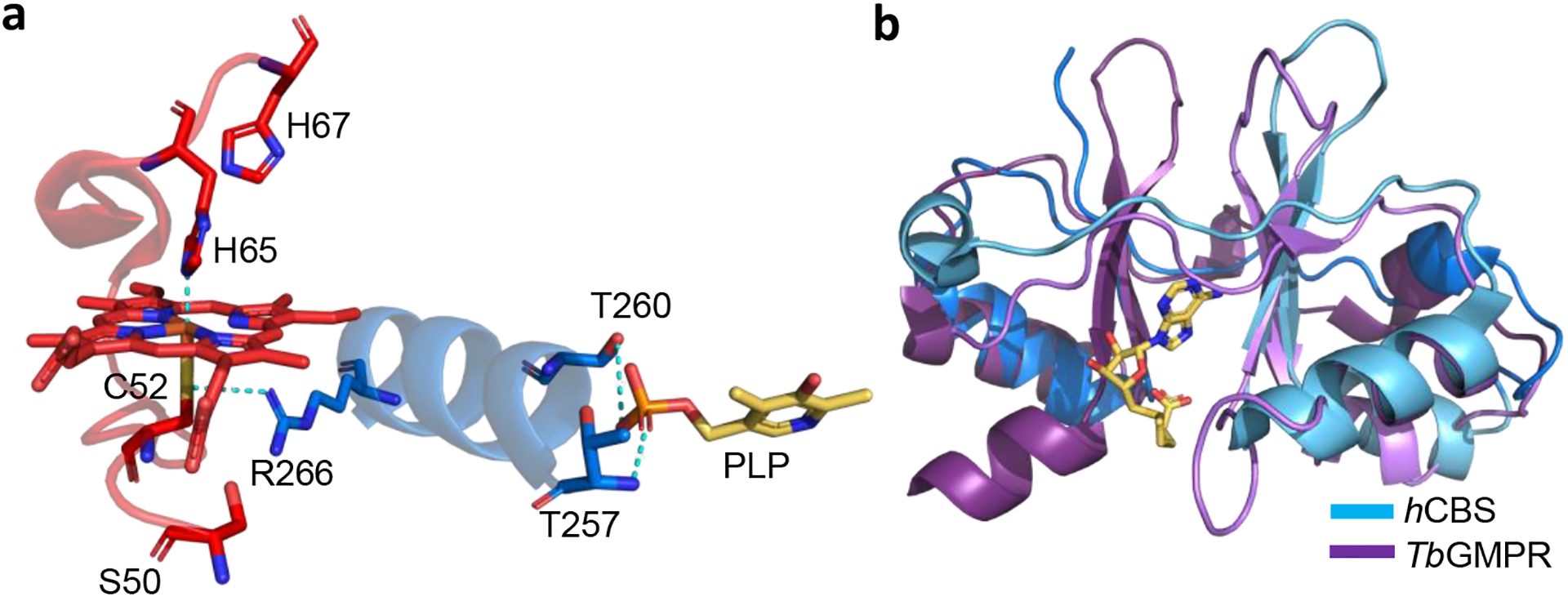
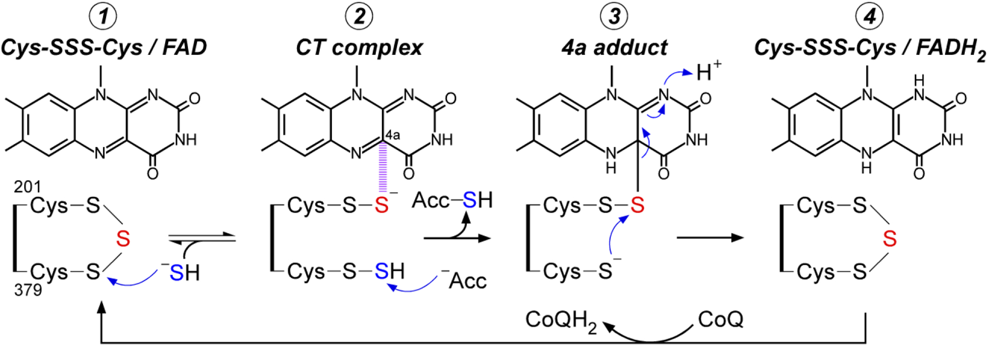
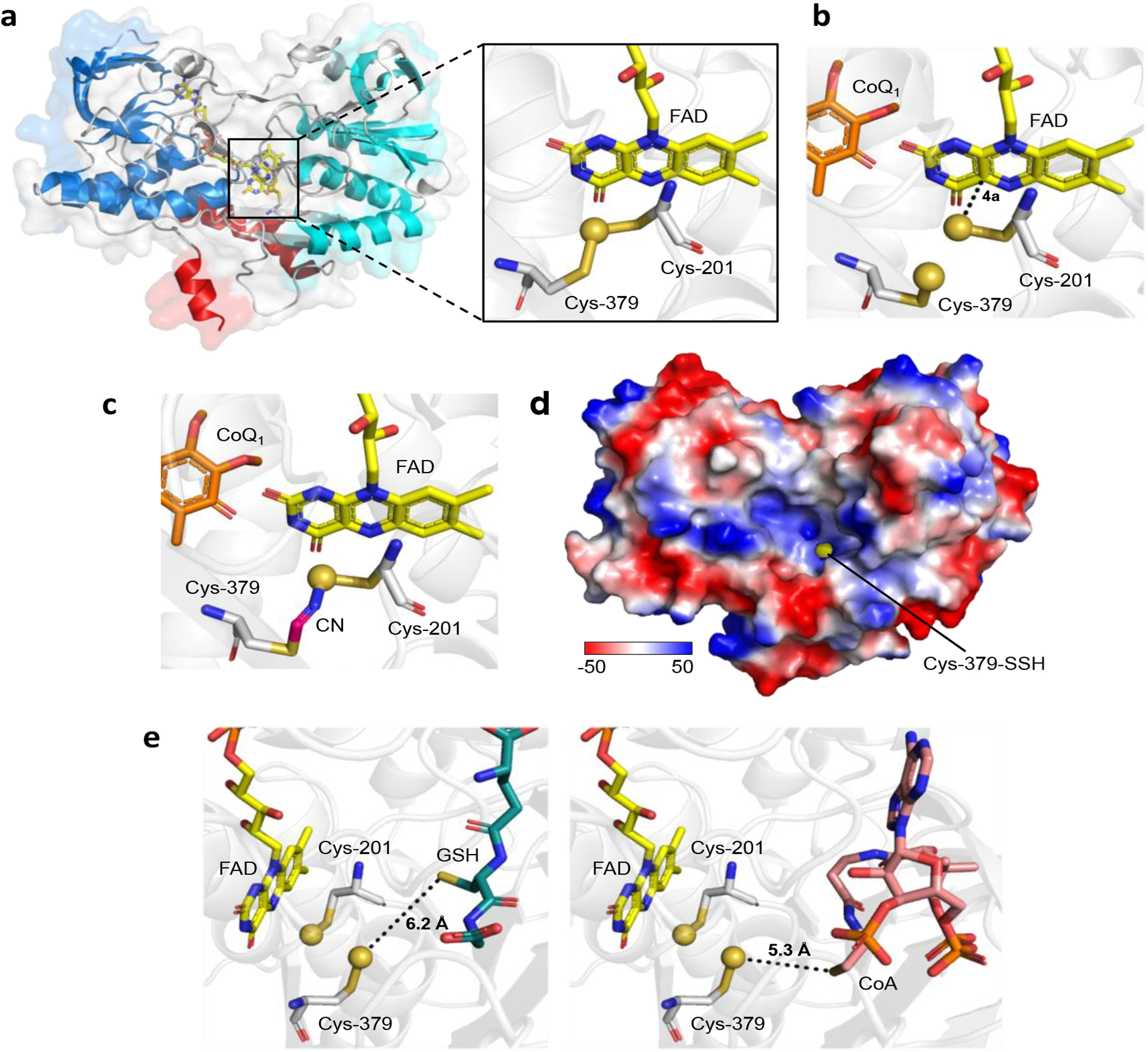
The enzymes involved in H2S homeostasis regulate its production from sulfur-containing amino acids and its oxidation to thiosulfate and sulfate. Two gatekeepers in this homeostatic circuit are cystathionine beta-synthase, which commits homocysteine to cysteine, and sulfide quinone oxidoreductase, which commits H2S to oxidation via a mitochondrial pathway. Inborn errors at either locus affect sulfur metabolism, increasing homocysteine-derived H2S synthesis in the case of CBS deficiency and reducing complex IV activity in the case of SQOR deficiency. In this review, we focus on structural perspectives on the reaction mechanisms and regulation of these two enzymes, which are key to understanding H2S homeostasis in health and its dysregulation and potential targeting in disease.


Conflict of interest statement

Conflict of interest statement R.B. is a paid member of the scientific advisory board of Apneo Therapeutics and owns equity in the company.

Figures

Figure 1.
Figure 1.. Enzymes involved in H2S homeostasis.
H2S biogenesis is catalyzed by three enzymes (blue): CBS, CSE and MPST while H2S catabolism is catalyzed by enzymes in the mitochondrial sulfide oxidation pathway (yellow). ETHE1, TST and SO denote persulfide dioxygenase, rhodanese and sulfite oxidase, respectively. CIII, CIV, 3-MP, Hcy and Cst denote complexes III and IV, 3-mercaptopyruvate, homocysteine and cystathionine, respectively.
Figure 2.
Figure 2.
Structure and functions of CBS. (a) Reactions catalyzed by CBS including the canonical transsulfuration reaction utilizing serine, the H2S generating reactions utilizing cysteine (± homocysteine) and the persulfide generating reaction utilizing cystine. (b) Domain organization of human CBS (upper) and the structures of CBS captured in a basal (PDB: 4COO, left) and an activated (PDB: 4PCU right) conformation. The domains are colored as in the key on top and CBS1 and CBS2 refer to the two CBS domains within the C-terminal regulatory segment that binds AdoMet.
Figure 3.
Figure 3.. CBS reaction mechanism and structure of two key intermediates.
(a) Postulated reaction mechanism for CBS-catalyzed H2O or H2S elimination. (b,c) Close-up of the structures of full-length Drosophila CBS in which a carbanion (PDB:3PC4) and an aminoacrylate (PDB: 3PC3) intermediate are seen. The residues and numbering in human CBS are shown in parentheses. Lys-88 (corresponding to Lys-119 in human CBS) is 2.5 Å from Cα providing electrostatic stabilization of the carbanion intermediate (b) but is rotated away and engaged in a hydrogen-bonding interaction with a phosphate oxygen in the aminoacrylate species (c). The PLP bound intermediates are shown in green.
Figure 4.
Figure 4.. Regulatory domains in CBS.
(a) Close-up showing the connection between the heme and PLP pockets in the structure of human CBS. Residues comprising the hydrophobic heme pocket are shown in red. His-65 and Cys-52 serve as the heme ligands while Cys-52 is also involved in an electrostatic interaction with Arg-266. (b) Overlay of the CBS domains in human CBS (PDB: 4PCU, blue) and T. brucei GMP reductase (PDB: 6JL8, purple). AdoMet bound to human CBS is shown in yellow stick display.
Figure 5.
Figure 5.. Proposed catalytic mechanism for SQOR.
The bridging sulfur of the cysteine trisulfide is shown in red and the sulfur undergoing oxidation is shown in blue. The dashed magenta lines denote a charge transfer (CT) complex between the Cys-201 persulfide and the flavin C4a, which leads to formation of the 4a adduct followed by FADH2.
Figure 6.
Figure 6.. Structure of human SQOR.
(a) Overall structure of native SQOR (PDB ID: 6OI5), with the first and second Rossmann fold domains shown in blue and cyan, respectively, and membrane-anchoring helices shown in in red. The inset is a close-up of the active site showing the cysteine trisulfide. (b) SQOR co-crystallized with CoQ (orange sticks; PDB ID: 6OIB), contains a persulfide pair, corresponding to [2] in Figure 5. (c) The SQOR Cys-379 N-(201Cys-disulfanyl)-methanimido thioate intermediate generated upon addition of cyanide (CN; PDB ID: 6WH6). (d) Electrostatic surface potential map showing the solvent exposure of Cys-379-SSH at the bottom of the electropositive cavity. (e) Models of physological acceptors docked in the SQOR cavity. The distances between the Cys-379 persulfide and the thiol moieties of GSH (left panel, teal sticks) and CoA (right panel, pink sticks) are noted. The sulfane sulfurs in all panels are shown as yellow spheres.

References

    1. Kabil O, Vitvitsky V, Banerjee R. Sulfur as a signaling nutrient through hydrogen sulfide. Ann Rev Nutr 2014; 34:171–205. - PMC - PubMed
    1. Chiku T, Padovani D, Zhu W et al. H2S biogenesis by human cystathionine γ-lyase leads to the novel sulfur metabolites lanthionine and homolanthionine and is responsive to the grade of hyperhomocysteinemia. J Biol Chem 2009; 284:11601–11612. - PMC - PubMed
    1. Singh S, Padovani D, Leslie RA et al. Relative contributions of cystathionine beta-synthase and gamma-cystathionase to H2S biogenesis via alternative trans-sulfuration reactions. J Biol Chem 2009; 284:22457–22466. - PMC - PubMed
    1. Yadav PK, Yamada K, Chiku T et al. Structure and kinetic analysis of H2S production by human mercaptopyruvate sulfurtransferase. J Biol Chem 2013; 288:20002–20013. - PMC - PubMed
    1. Yadav PK, Vitvitsky V, Carballal S et al. Thioredoxin regulates human mercaptopyruvate sulfurtransferase at physiologically-relevant concentrations. J Biol Chem 2020; 295:6299–6311. - PMC - PubMed

Publication types

LinkOut - more resources